
Fig. 5
|
Fig. 5
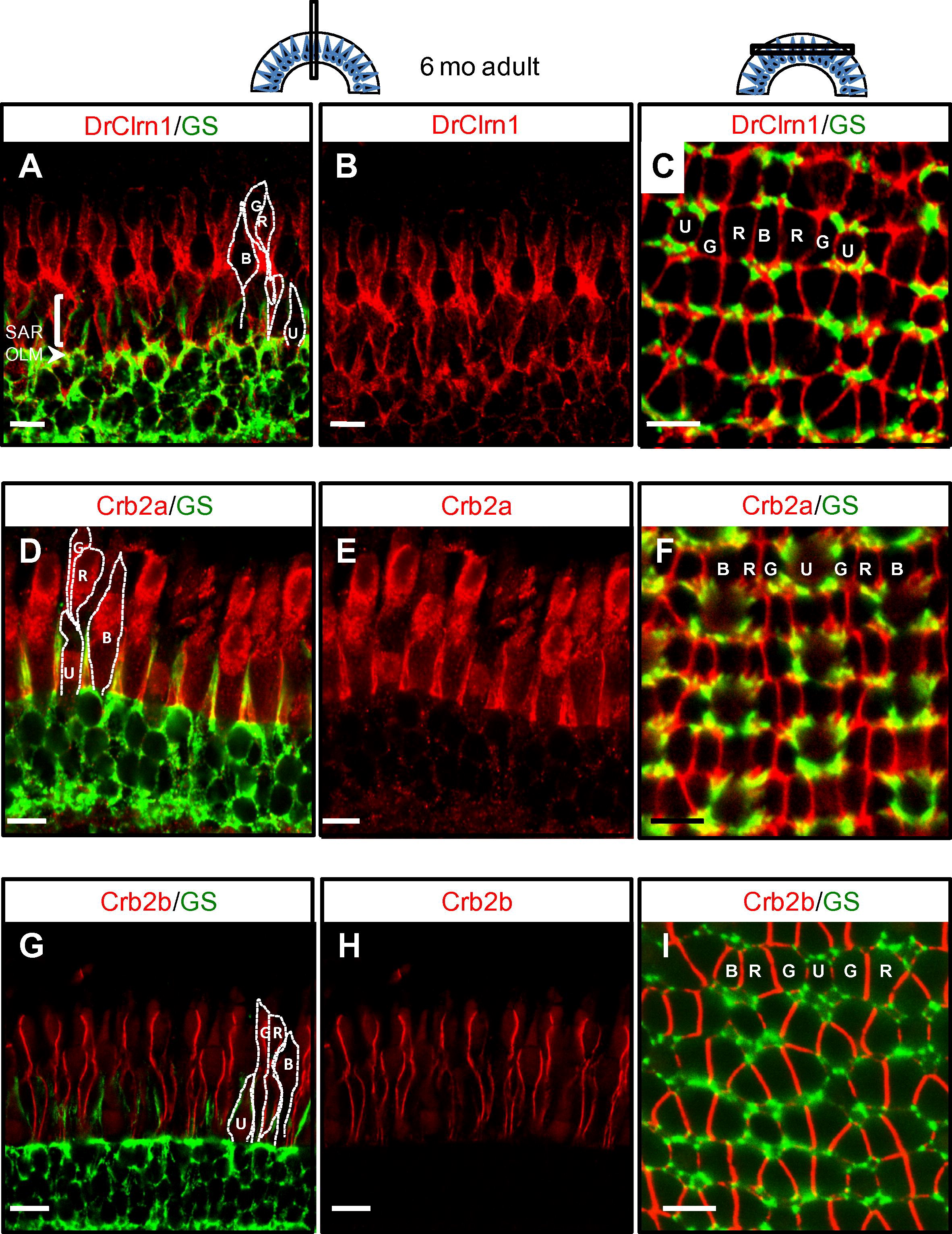
Clarin-1 localizes at interfaces between photoreceptor inner segments in the adult retina. (A-C) Cross (A and B) and transverse (C) sections through adult retinas show Clarin-1 localization partially overlapping with glial cells labeled with Glutamine Synthetase (GS) at the OLM (arrowhead). Enrichment of Clarin-1 is also noted in the subapical region (SAR) and between all cone inner segments in the outer retina. The silhouettes of the four cone subtypes are outlined in white in A, D and G and the mosaic cone arrangement is labeled in C, F and I. (D-I) Cross (D-E, G-H) and transverse (F, I) sections of adult retinas labeled with Crb2a (D-F) and Crb2b (G-I) antibodies recapitulate results published by Zou et al., 2012, and provide points of reference for the Clarin-1 (A-C) localization pattern at lateral cone interfaces. Abbreviations: OLM: outer limiting membrane; G: green opsin cone, R: red opsin cone; B: blue opsin cone; SAR (subapical region) U: UV opsin cone. Scale bars: 10 µm.
Reprinted from Gene expression patterns : GEP, 13(8), Phillips, J.B., Västinsalo, H., Wegner, J., Clément, A., Sankila, E.M., and Westerfield, M., The cone-dominant retina and the inner ear of zebrafish express the ortholog of CLRN1, the causative gene of human Usher syndrome type 3A, 473-81, Copyright (2013) with permission from Elsevier. Full text @ Gene Expr. Patterns