
|
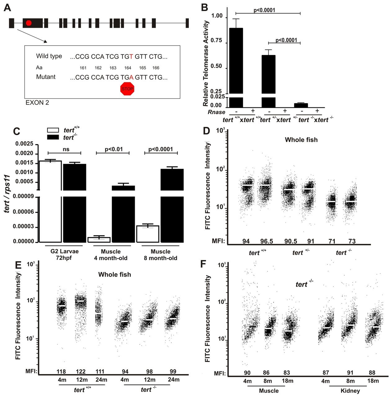
Fig. 1
The dynamics of telomere and telomerase in tert mutant zebrafish. (A) Schematic representation of the telomerase gene mutation. (B) Telomerase activity was measured quantitatively in whole zebrafish embryos (3 dpf, n=100) by Q-TRAP using 0.1 mg of protein extract. Results are expressed as the mean value ± s.e.m. from triplicate samples relative to telomerase-positive cells. Statistical significance was assessed using Student’s t-test (P<0.05). To confirm the specificity of the assay, a negative control is included for each sample, treated with 1 µg of RNase at 37°C for 20 minutes. (C) The mRNA levels of tert gene were determined by real-time RT-PCR in larvae and adult muscle tissue of the indicated genotypes. Gene expression is normalized against rps11. Each bar represents the mean ± s.e.m. from 100 pooled animals for larvae and three individual fish for adult tissue and triplicate samples. (D) Representation of 3-month-old zebrafish cell distribution according to telomere length. Medium fluorescence intensity (MFI) is indicated for each genetic background. The same trend was observed in the three independent experiments. (E) Representation of wild-type and tert mutant zebrafish cell distribution throughout their life according to their telomere length. MFI is indicated for each genetic background. The same trend was observed in the three independent experiments. m, months. (F) Representation of zebrafish muscle and kidney cell distribution throughout life according to their telomere length. MFI is indicated for each genetic background. The same trend was observed in the three independent experiments.