
|
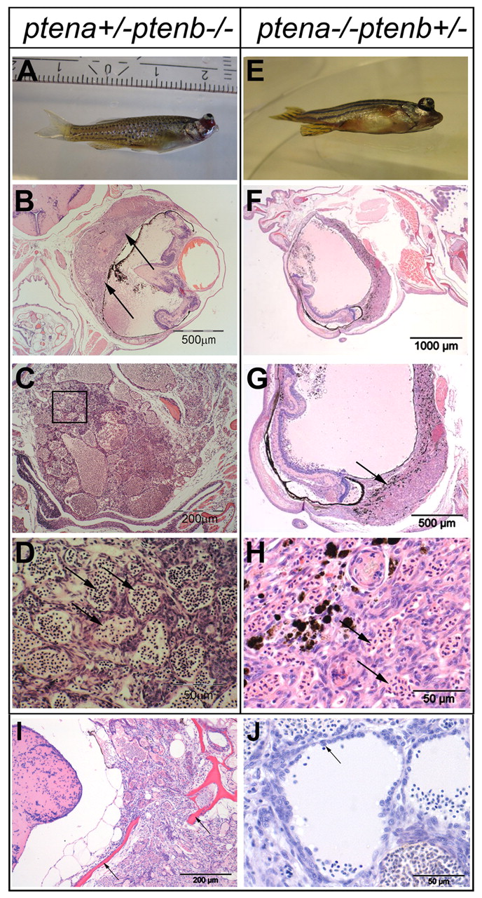
Fig. 2
Ocular tumor of ptena +/-ptenb -/- and ptena -/-ptenb+/- mutants, diagnosed as hemangiosarcoma. (A-D) 3-month-old ptena+/-ptenb -/- and (E-H) 9-month-old ptena -/-ptenb+/- mutant with ocular tumor. The entire intact fish was fixed and embedded in paraffin. (B-D) Transversal sections and (F-H) sagittal sections were stained with H&E. Arrows indicate tumor mass, which is associated with the eye bulbs. (B,G) Higher-power magnifications of the tumor mass; (D) magnification of the boxed area in C. The tumor consists of cells that form different sizes of blood-filled spaces (arrows in D,H).(I,J) H&E staining of sections from two individuals revealed hemangiosarcoma formation. (I) The tumor was invasive and penetrated into the brain region with enclosing scull elements (arrows). (J) Cells with plump morphology (arrow) are detaching from surrounding tissue and protrude into the vessel lumen. Sections of representative tumors are depicted here.