
|
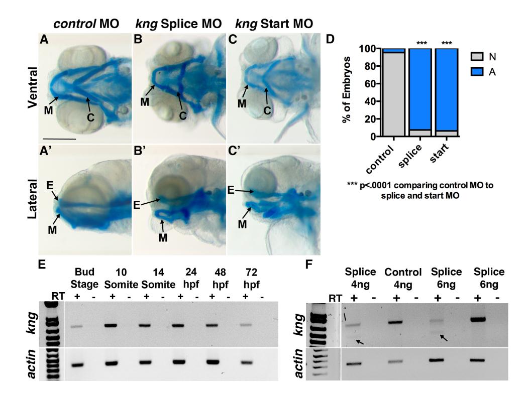
Fig. S6
kininogen (kng) is expressed throughout zebrafish craniofacial development and loss of function results in craniofacial cartilage abnormalities, related to Figure 7.
(A-C, A′-C′) kng loss of function using splice site and start site morpholinos. Embryonic cartilage was observed at 5dpf using alcian blue staining in three independent experiments. E, Ethmoid plate. C, Ceratohyal cartilage. M, Meckel′s cartilage. Scale bar: 250µm. (A,A′) Control morpholino injected embryos appeared normal (95% normal, n=43). (B-B′, C-C′) Splice and start morphants showed abnormal facial cartilage. Meckel′s cartilage is truncated and lies at an abnormal angle. The ceratohyal cartilage points at an abnormal angle, perpendicular to the midline. (kng splice morpholino (B,B′) 7% normal, n=95; kng start morpholino (C,C′) 6% normal, n=47). (D) Quantification of morphant phenotypes. P-values: one-tailed Fisher Exact test. (E) Non-quantitative RT-PCR. kng expression is present at bud stage and extends until 72hpf. Expression spans mouth opening at 48hpf and formation of facial cartilage at 72hpf. (F) Non-quantitative RT-PCR. RNA was extracted at 24hpf. Controls yield normal length kng transcripts, while splice morphants yield fewer normal length transcripts and a truncated transcript. Arrow: truncated transcript band.