
Image
|
Figure Caption
Fig. S6
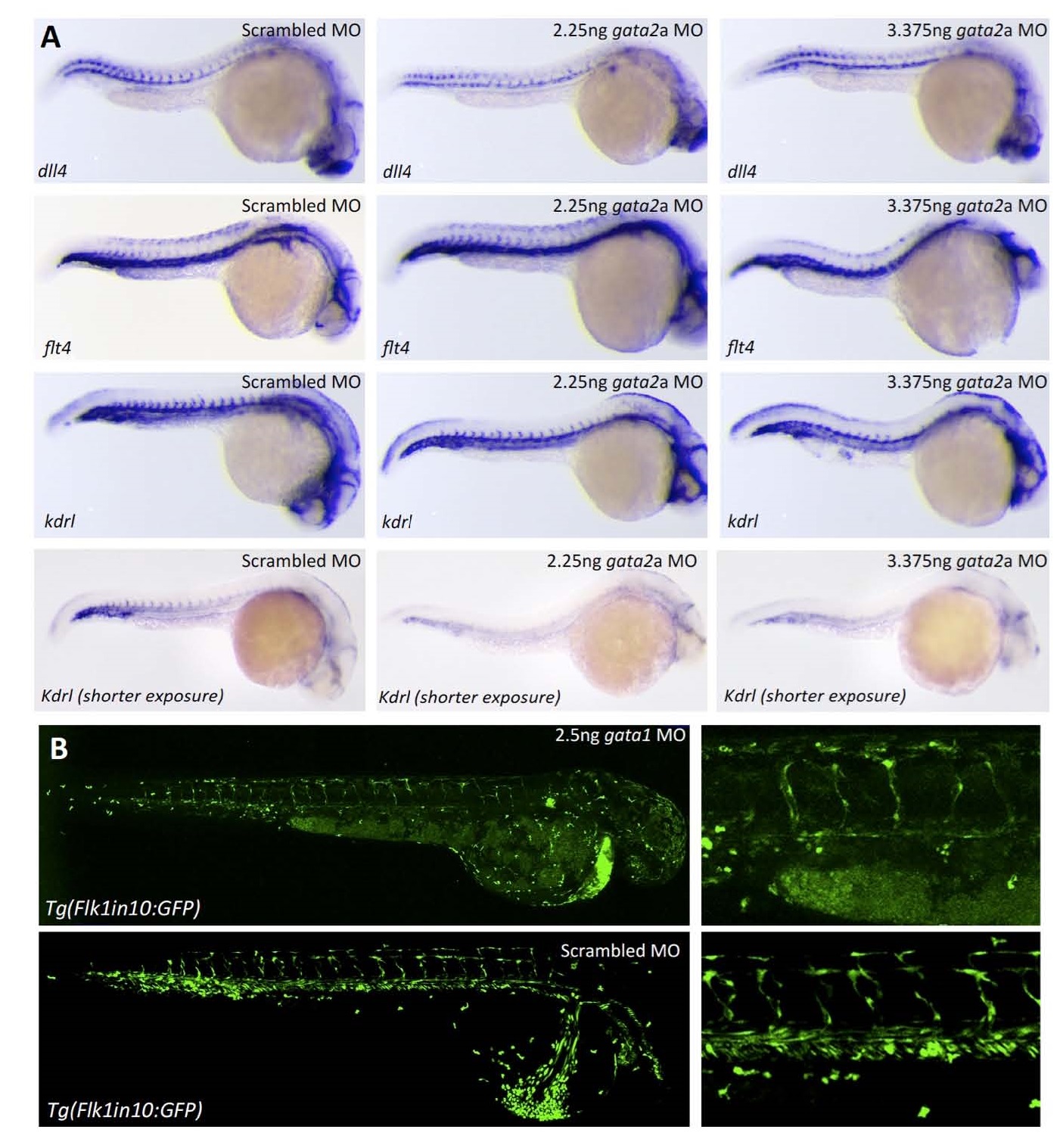
A. Analysis of scrambled, 2.25ng and 3.375 ng gata2a MO in 26 hpf WT zebrafish embryos, using whole-mount in situ hybridization with probes against arterial marker dll4, venous marker flt4 and kdrl.
B. Analysis of 2.5ng gata1 MO in 36 hpf tg(Flk1in10:GFP) embryos.
Acknowledgments
This image is the copyrighted work of the attributed author or publisher, and
ZFIN has permission only to display this image to its users.
Additional permissions should be obtained from the applicable author or publisher of the image.
Full text @ Arterio., Thromb., and Vas. Bio.