
|
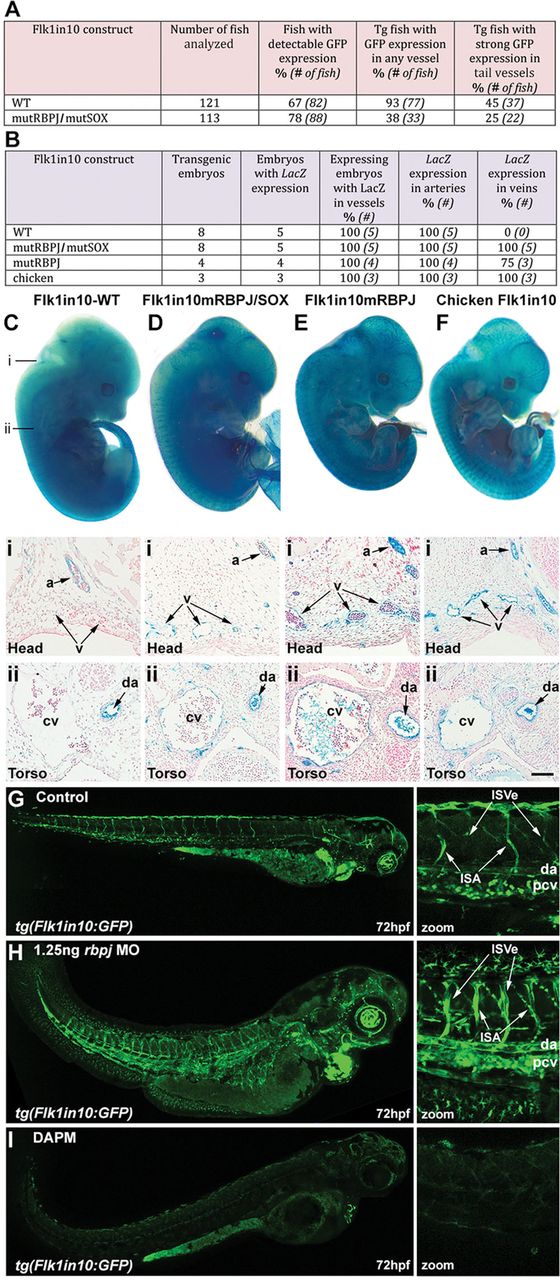
Fig. 5
Rbpj is required for arterial restriction of the Flk1in10 enhancer. A and B, Summary of reporter gene expression detected in (A) 48 h post fertilization (hpf) Tol2-mediated mosaic transient transgenic zebrafish embryos and (B) embryonic day (E) 12 transient transgenic mice. C-F, Whole-mount (top) and transverse sections (bottom) of representative E12 X-gal-stained transient transgenic embryos expressing mouse Flk1in10 WT (C), mouse Flk1in10mRBPJ/SOX (D), mouse Flk1in10mRBPJ (E), and chicken Flk1in10 WT (F). Lines marked i and ii on C mark the approximate location of transverse sections in C-F. a indicates artery; cv, cardinal vein; da, dorsal aorta; and v, veins. G-I, Analysis of the effects of scrambled (G), 1.25 ng rbpj morpholino (MO) oligonucleotides (H), and Notch signaling inhibitor N-[N-3,5-difluorophenacetyl]-l-alanyl-S-phenylglycine methyl ester (DAPM) (I) in 72 h post fertilization (hpf) tg(Flk1in10:GFP) embryos. MO-mediated knockdown of rbpj results in expansion of Flk1in10:GFP expression into the caudal vein, the posterior cardinal vein, and intersegmental vein. da indicates dorsal aorta; GFP, green fluorescent protein; ISA, intersegmental artery; ISVe, intersegmental vein; and pcv, posterior cardinal vein.