
Image
|
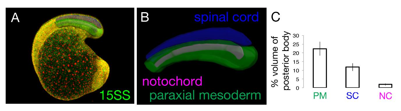
Figure Caption
Fig. S1
Proportional volumes of the spinal cord, paraxial mesoderm and notochord with respect to the whole posterior body. (A,B) Confocal stacks of 15-somite stage embryos injected at the one-cell stage with nuclear mCherry and membrane-bound eGFP mRNAs were used to build surface reconstructions of the spinal cord (blue), paraxial mesoderm (green) and notochord (purple). (C) Proportional volume of each tissue with respect to whole posterior body volume. Measurements are from three embryos.
Acknowledgments
This image is the copyrighted work of the attributed author or publisher, and
ZFIN has permission only to display this image to its users.
Additional permissions should be obtained from the applicable author or publisher of the image.
Full text @ Development