
|
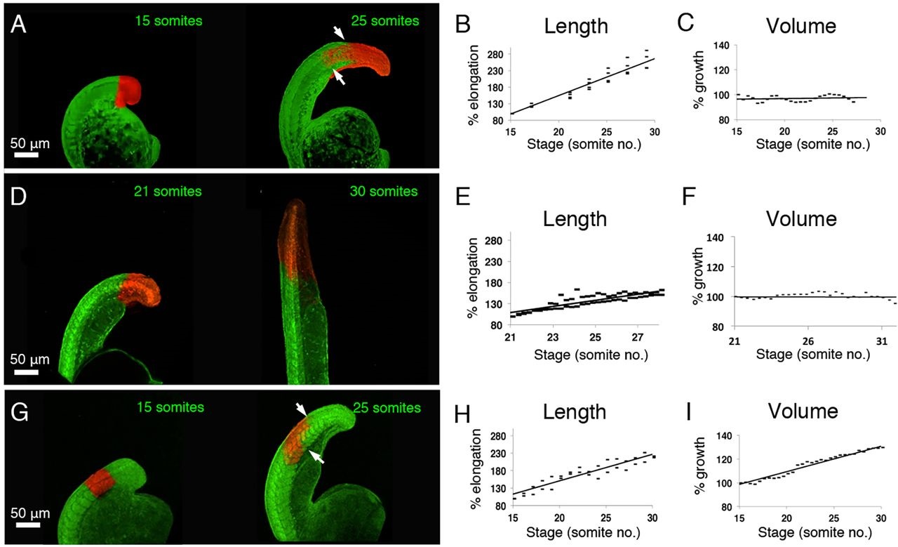
Fig. 3
Volumetric growth only occurs within already segmented regions of the zebrafish posterior body. (A,D,G) Stills from time-lapse movies of embryos injected at the one-cell stage with KikGR mRNA and photolabelled within the unsegmented region at the 15-somite (A) or 21-somite (D) stage or within the segmented region at the 15-somite stage (G). Embryos in lateral view with posterior to the top. (B,E,H) Photoactivated region length (percentage of initial length) plotted against time (number of somites). (B) n=25, (E) n=51, (I) n=29. (C,F,I). Photoactivated region volume (percentage of initial volume) plotted against time (number of somites), (C) n=25, (F) n=25, (I) n=31. White arrows in A and G indicate the displacement of labels within the somitic mesoderm (lower arrows) relative to the spinal cord (upper arrows). Data points show individual measurements; n=total number of measurements from three independent experiments.