
Fig. 4
- ID
- ZDB-IMAGE-160524-63
- Publication
- Trowe et al., 1996 - Mutations disrupting the ordering and topographic mapping of axons in the retinotectal projection of the zebrafish, Danio rerio
- All Figures
- Figures for Trowe et al., 1996
|
Fig. 4
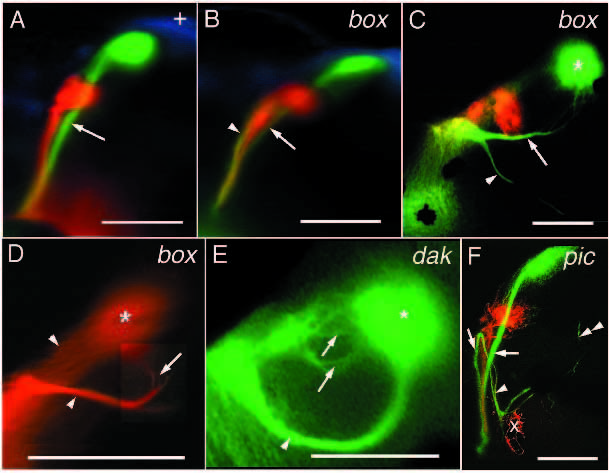
Mutations affecting the sorting of axons into the two branches of the optic tract. (A) In wild-type animals, nasodorsal axons (green) grow in the ventral branch of the optic tract (arrow). (B) In box mutant fish, nasodorsal axons grow in the ventral (arrow) and in the dorsal branch (arrowhead) of the optic tract. (C) Another box mutant fish. Nasodorsal axons that enter the tectum through the dorsal branch continue their dorsal trajectory on the tectal lobe (arrow). Near the posterior margin of the lobe, they turn and terminate retinotopically, posteroventrally (*). Some of the ectopic nasodorsal axons leave the contralateral tectal lobe and grow to the ipsilateral one (arrowhead). (D) Trajectory of middorsal RGC axons of a box mutant. Dorsal view of the contralateral tectal lobe. Middorsal axons grow in both branches of the optic tract and on the dorsal and the ventral side of the tectal lobe (arrowheads). Ectopic middorsal axons make a sharp turn middorsally (arrow) and grow to their retinotopic midventral target area (*). (E) Nasodorsal axons in a dak mutant. As in box, these axons grow in both branches of the optic tract. Ectopic nasodorsal axons grow around the dorsal side of the tectal lobe (arrowhead), but terminate retinotopically, posteroventrally (*). The nasodorsal axons on the dorsal side of the lobe are much more strongly fasciculated than the ones on the ventral side (arrows). (F) Confocal image of the retinotectal projection of a pic mutant. Nasodorsal axons grow in both branches of the optic tract (arrows). The nasodorsal axons in the dorsal branch and some temporoventral axons turn and grow to the ipsilateral tectal lobe (arrowhead). Here, the temporoventral axons terminate retinotopically (x), while the nasodorsal axons first grow around the dorsal margin of the lobe and then leave it to grow towards the contralateral lobe again (double arrowhead). Scale bars, 0.1 mm.