
|
Fig. 1
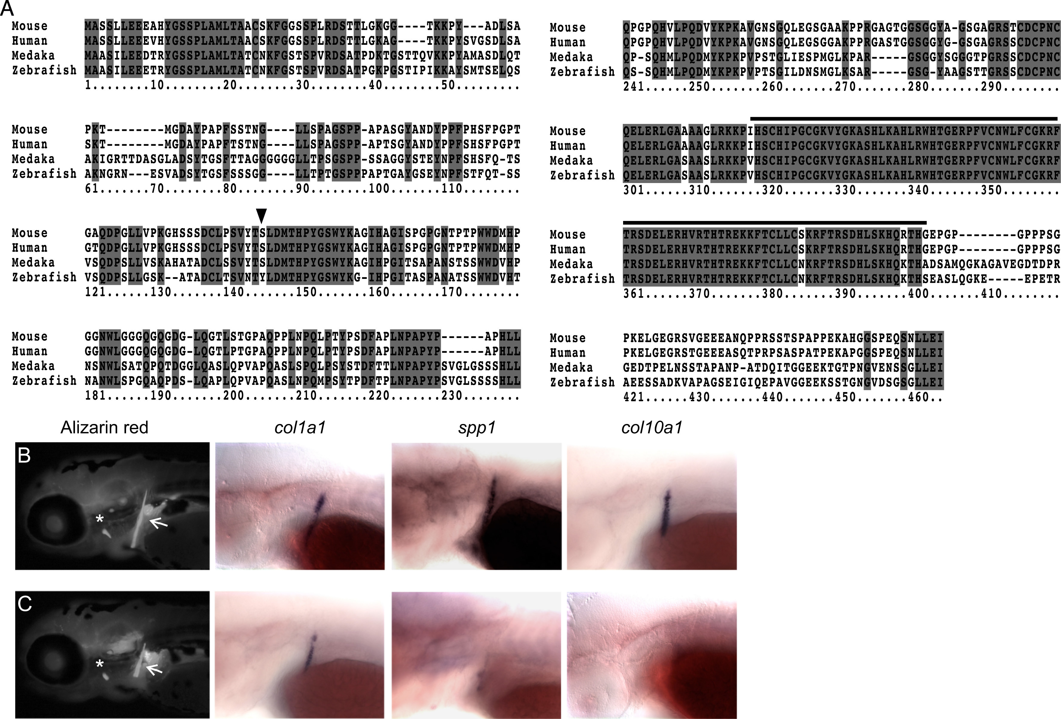
Zebrafish sp7 mutants have delayed early bone development. (A) Sp7 protein sequences of human, mouse, medaka and zebrafish were aligned using ClustalW. Identical residues are shaded dark gray. Cysteine and histidine residues corresponding to the zinc finger domains from positions 318 to 402 (black line) are highly conserved in a domain showing only three amino acid variations between mammals and fish. The mutation p.Leu145* (black triangle) introduces a stop codon, deleting over half of the Sp7 coding sequence, including the three zinc finger domains. (B and C). Calcified bones were detected at 4 dpf by vital Alizarin red staining. While WT (B) and mutant (C) larvae were indistinguishable by morphology, the cleithrum (arrows) and opercle (asterisks) are slightly smaller in sp7 mutants. In situ hybridization was performed for markers of osteoblast differentiation in embryos at 4 dpf. Expression of all markers in the cleithrum was reduced in mutants, especially spp1 and col10a1.
Reprinted from Developmental Biology, 413(2), Kague, E., Roy, P., Asselin, G., Hu, G., Stanley, A., Albertson, C., Simonet, J., Fisher, S., Osterix/sp7 limits cranial bone initiation sites and is required for formation of sutures, 160-72, Copyright (2016) with permission from Elsevier. Full text @ Dev. Biol.