
|
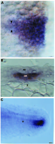
Fig. 4
Relationship of rtk1 expression to the developing notochord. Whole-mounted embryos labelled with an antisense RNA probe to rtk1 (A-C) and antibody to Ntl (A,B). Anterior is to the left in A and C and dorsal is up in B and C. (A) 60% epiboly. rtk1 transcripts (blue) and Ntl protein (brown) are colocalised at the shield, although rtk1 expression is detectable along the axis slightly anterior (arrowheads) to Ntl-containing cells. Ntl is also within germ ring cells lateral to the shield. (B) Transverse section through the presumptive notochord. rtk1 is expressed in the same cells that contain Ntl protein. (C) rtk1 expression is downregulated as the notochord differentiates but continues to be expressed in presumptive notochord cells of the extending tail bud. Abbreviations: n, notochord, ne, neuroepithelium; pn, presumptive notochord; y, yolk. Scale bars: 25 µm.