
|
Fig. 5
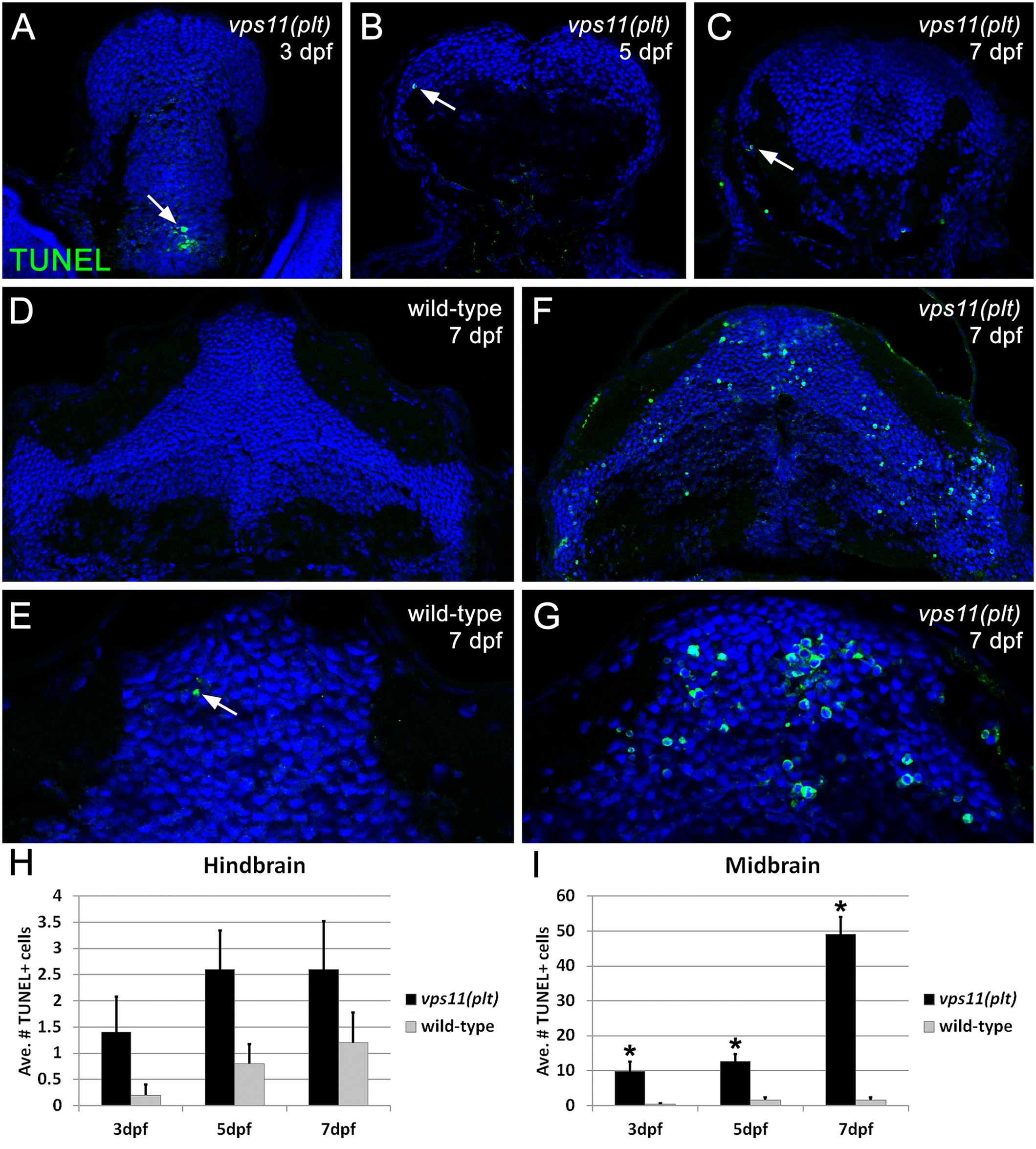
Significant cell death is observed in the CNS of zebrafish vps11(plt) mutants.TUNEL assay was performed on cryosectioned CNS tissue from vps11(plt) mutants and wild-type siblings at 3, 5, and 7 days post-fertilization (dpf). (A-C) Minimal apoptotic TUNEL+ cells were observed in the hindbrain of vps11(plt) mutants at 3, 5, and 7 dpf, respectively. (D-E) Tissue section of the midbrain of a wild-type control animal at 7 dpf showing minimal cell death (panel E, arrow). (F-G) Tissue section of the midbrain of a vps11(plt) mutant at 7 dpf showing extensive cell death. (H) Quantification of the average number of TUNEL+ cells observed in the hindbrain in vps11(plt) mutants and wild-type siblings at 3, 5, and 7 dpf. No significant differences were observed. (I) Quantification of the average number of TUNEL+ cells observed in the midbrain in vps11(plt) mutants and wild-type siblings at 3, 5, and 7 dpf. Asterisk indicates significantly different from control.