
|
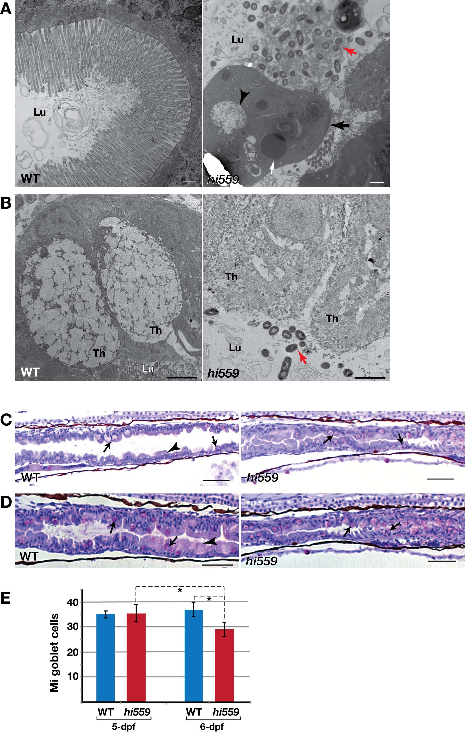
Fig. S2
Abnormal goblet cells and mucosecretion in hi559 intestinal mucosa. (A) Wild-type intestinal luminal area appears clear with mucinous granules and microvilli projections from IECs. The hi559 lumen is often filled with detached cells (black arrow) and bacterial overgrowth at 5.5-dpf (red arrow). Nuclear condensation (white arrow) and cytoplasmic vacuoles (arrowhead) are apparent in the detached IEC, suggesting apoptosis. (B) At 5-dpf, wild-type mucinous GCs are mature with largely dense mucinous theca, whereas the hi559 GCs have poorly developed theca with abnormal vesicles, cytoplasmic necrosis and bacterial invasion (red arrow). (C-D) PASstaining (pink) showing the mucin-secreting mid-intestinal GCs (arrow) and secreted mucinous layer (arrowhead) of the mid-intestinal epithelium at (C) 5-dpf and (D) 6-dpf. The thick secreted mucinous layer, which is seen at the apical border of wild-type intestinal mucosa, is greatly diminished in hi559 and the mucinous cells are disarranged and often detached into the lumen. (E) Bar-chart showing the number of mid-intestinal (Mi) GCs in wild-type and hi559 intestine at 5 and 6-dpf (n=7, *P<0.05). Goblet cell population is significantly depleted in hi559 intestine at 6-dpf. Wild-type (WT) on left and hi559 on right panels. Lu, intestinal lumen; Th, mucinous theca. Scale bar: A-B, 500 nm; C-D, 20 µM.