
|
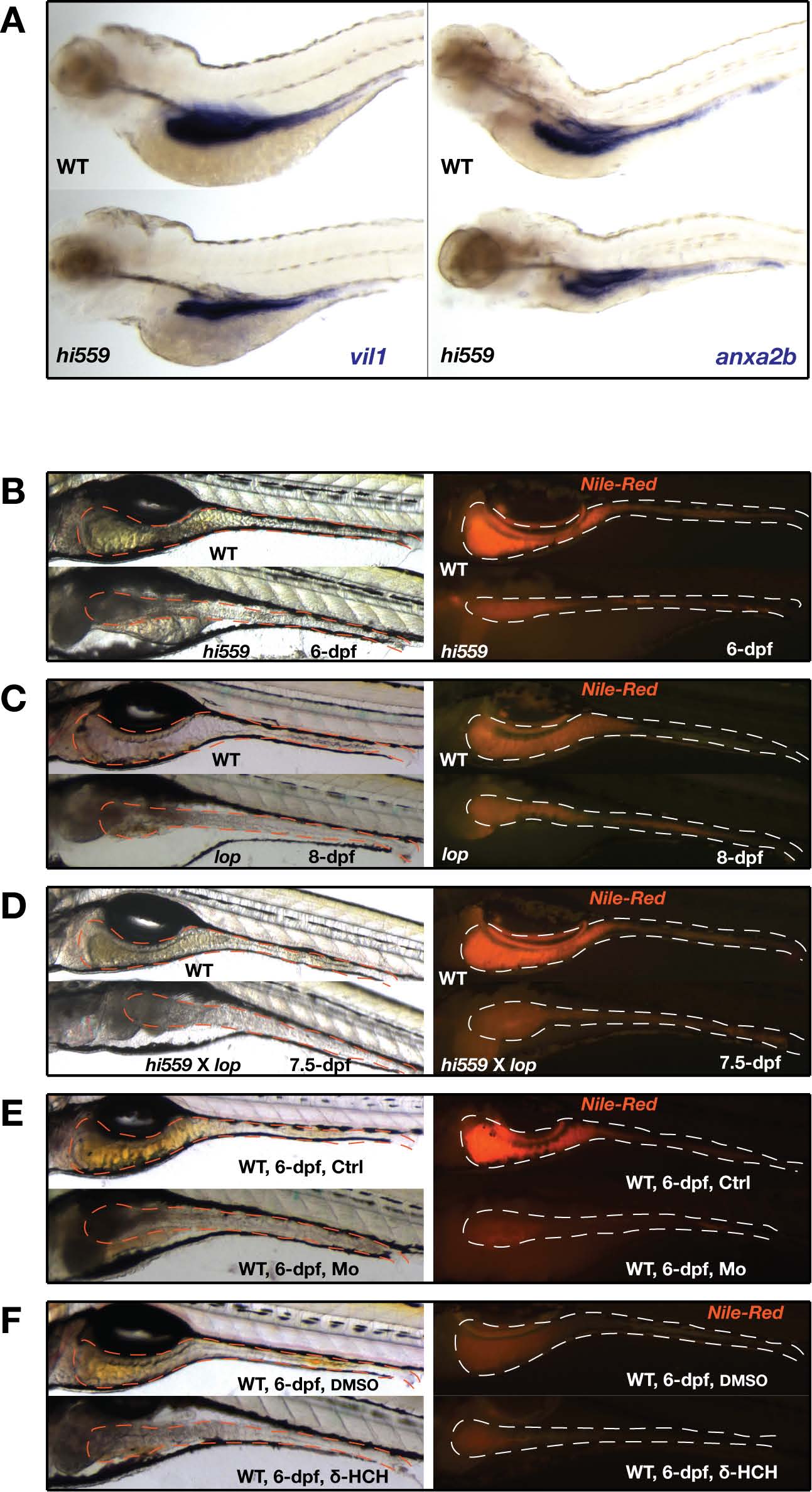
Fig. S1
The cdiptlop (lop) and cdipthi559 (hi559) mutants exhibit identical GI phenotype and fail to rescue each other in complementation assay. (A) Lateral view of ISH of intestine-specific markers vil1 and anxa2b showing reduced expression in the hi559 intestine at 5-dpf. (B-F) Morphology of intestine shown by bright-field images (left panel) and fluorescent images (right panel, Nile-Red staining shown by white outline). (B) GI morphology of hi559 larvae at 6-dpf shows smaller intestine and lumen. (C) GI morphology of lop larvae shows smaller intestine and lumen at 8-dpf comparable to the 6-dpf hi559 larvae. (D) Cdipthi559/lop (hi559 X lop) trans-heterozygote mutant larvae show smaller intestinal lumen at 7.5-dpf comparable to the 6- dpf hi559 larvae. (E) Wild-type embryos were microinjected with either control morpholino or splice-blocking morpholino against cdipt at 1-cell stage and larvae were analyzed at 6-dpf (Thakur et al., 2011). Cdipt morphants show smaller intestinal structures replicating hi559 phenotype. (F) Wild-type larvae were treated with PI synthase inhibitor δ-HCH shows smaller intestinal structures compared to DMSO treated larvae.