
|
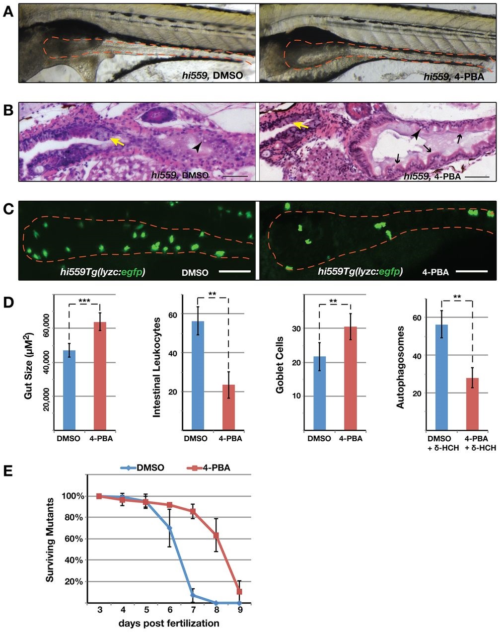
Fig. 7
Chemical chaperone 4-PBA rescues the hi559 GI phenotype. (A) Bright-field image of DMSO- and 4-PBA-treated hi559 larvae at 6.5 dpf, showing amelioration of gross intestinal structure (red outline). (B) H&E-stained sagittal sections shows improved villous architecture (black arrows), GCs (yellow arrows) and reduction in mucosal necrosis (arrowheads) in 4-PBA-treated larvae. (C) Confocal projection of hi559Tg(lyzc:egfp) larval intestine (red outline) showing reduction in intestinal leukocyte infiltration in 4-PBA-treated larvae. (D) Bar charts of hi559 larval gut size (n=15), intestinal leukocyte counts in hi559Tg(lyzc:egfp) larvae (n=12) and intestinal autophagosomes (lc3:gfp punctates) in δ-HCH-treated wild-type Tg(lc3:gfp) larvae (n=7) exposed to DMSO or 4-PBA. (E) Survival curve of hi559 larvae treated with DMSO or 4-PBA from 3 dpf through to 9 dpf. Error bars indicate s.d. **P<0.01, ***P<0.001. Scale bars: 20 µm (B); 50 µm (C).