
|
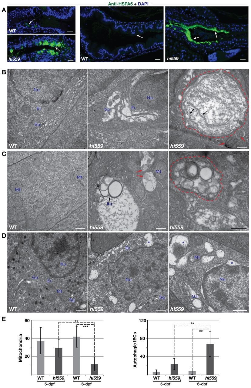
Fig. 4
ER stress and ultrastructural pathology of IECs. (A) Anti-HSPA5 immunofluorescence assay (green) shows robust enrichment of Hspa5 protein in the GCs (arrows, left panel) and the IECs along the epithelial lining (arrows, right panel) of hi559 intestine compared with wild type (WT). (B-D) TEM comparison of wild type (left panels) and hi559 IECs (middle and right panels). (B) ER-Golgi compartments are grossly expanded in 5-dpf hi559 IECs. Large double-membranous autophagic vacoules (red outline) and pre-autophagosome structures (red arrows), containing ER fragments (black arrows) are apparent in 5.5-dpf hi559 IECs (right panel). (C) Wild-type IECs have abundant mitochondria, whereas hi559 IECs have depleted, abnormal mitochondria and increased mitophagy at 6 dpf (red arrows). Multi-lamellar autophagic bodies (red outline), engulfing organelles, occur frequently in hi559 IECs at 6 dpf. (D) Secretory granule-rich enteroendocrine cells show ER luminal swelling (asterisks) and autophagic vesicles in hi559. Nu, nucleus; Er: endoplasmic reticulum; Ga, Golgi apparatus; Au, autophagosome, Sg, secretory granules. Mt, mitochondria. (E) Bar charts of mitochondrial (left) and autophagosome (right) counts in IECs (n=7); **P<0.01, ***P<0.001. Scale bars: 20 µm (A); 500 nm (B-D).