
|
Fig. 1
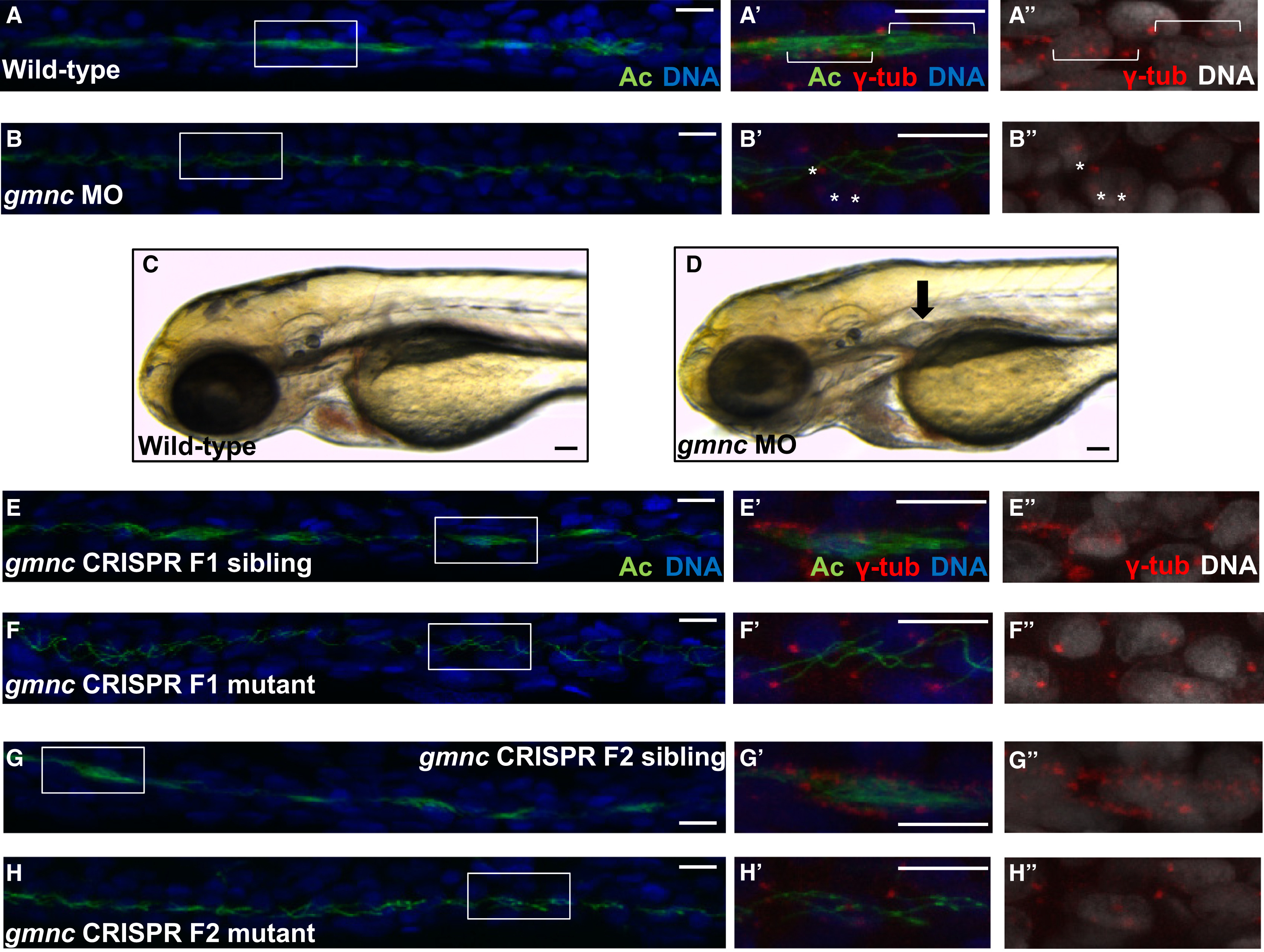
Gmnc Is Required for MCC Formation in the Kidney Tubules of the Zebrafish Embryo
(A and B) The kidney tubules of 48 hr post-fertilization (hpf) wild-type embryos are populated with clusters of MCCs (A). Boxes delineate the regions imaged at a higher magnification in (A′) and (A′′). Multiple basal bodies are indicated by brackets (A′ and A′′). In contrast, 100% (n = 47, from two independent experiments) of the gmnc splice-blocking MO-injected embryos were devoid of MCCs (B and B′). Examples of single basal bodies are marked by asterisks (B′ and B′′). Acetylated-tubulin (Ac; green), DAPI (DNA; blue; rendered in black and white in A′′ and B′′), and γ-tubulin (marks basal bodies; red; for A′, A′′, B′, and B′′ only).
(C and D) In contrast to wild-type embryos (C), kidney cysts (black arrow) form in 3-day-old gmnc morphants (93%, n = 40) (D).
(E and F) The F1 generation from intercrossed F0 gmnc CRISPR mutants was a mixture of wild-type animals (E) and animals carrying the gmnc deletion (F). The kidney tubules of the latter did not harbor MCCs at 36 hpf.
(G and H) Approximately one-quarter (two different pairs of F1 heterozygotes were analyzed; pair 1: 26%, n = 31; pair 2: 27%, n = 44) of the F2 generation from intercrossed heterozygous F1 parents lacked MCCs in the kidney tubules at 36 hpf (H). The other three-quarters of the embryos appeared wild-type (G).
Scale bars, 10 µm (C and D); 5 µm (all others). See also Figure S1.