
|
Fig. 4
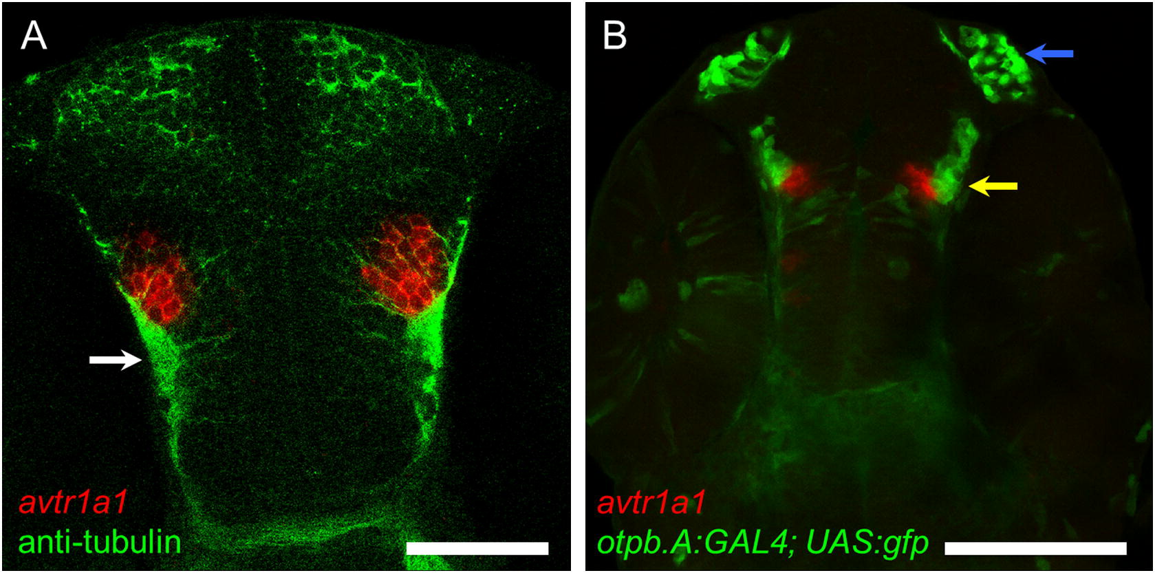
At 48 hpf the avtr1a1+ cells in the anterior forebrain (I) are the POC/TPOC neurons and not preoptic neurons. (A) Dorsal perspective (anterior up) of an embryo labeled with avtr1a1 riboprobe (red) and anti-acetylated α tubulin labeled axons (green) showing that the forebrain avtr1a1+ cells appear to extend axons in the TPOC (arrow). Scale: 50 µm. (B) A ventral perspective of an otpb.A:GAL4; UAS:gfp embryo labeled with avtr1a1 riboprobe showing that the anterior forebrain avtr1a1+ neurons (red) are located just medial to the preoptic neurons (green, yellow arrow). Blue arrow denotes the olfactory neurons that express GFP in the transgenic embryos. Scale: 100 µm.
Reprinted from Gene expression patterns : GEP, 13(8), Iwasaki, K., Taguchi, M., Bonkowsky, J.L., and Kuwada, J.Y., Expression of Arginine Vasotocin Receptors in the Developing Zebrafish CNS, 335-42, Copyright (2013) with permission from Elsevier. Full text @ Gene Expr. Patterns