
|
Fig. 1
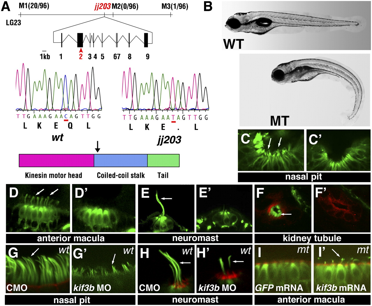
The jj203 mutant locus encodes the kif3b kinesin subunit. (A) Positional cloning of jj203 mutant locus. Top: Map of the jj203 genomic region and exon/intron structure of the kif3b transcript. Middle: Sequence of the jj203 transcript in WT and mutant animals. Bottom: Approximate diagram of Kif3b protein domain structure. Arrow indicates the site of the stop codon in the jj203 mutant allele. (B) The external phenotype of WT (Upper) and jj203 mutant (Lower) larvae at 5 dpf. (C-F′) Confocal images of WT (C, D, E, and F) and jj203 mutant (C′, D′, E′, and F′) larvae immunostained to visualize ciliogenesis in olfactory epithelium (C and C′), auditory maculae (D and D′), lateral line neuromasts (E and E′), and kidney tubules (F and F′) at 2 dpf (C and C′) or 3 dpf (D-F′). C-E′ show images of whole animals; F and F′ show transverse cryosections. (G-H′) Confocal images of olfactory pits (G and G′) and lateral line neuromasts (H and H′) in zebrafish larvae treated with anti-kif3b or with a control (CMO) antisense morpholino as indicated. (I and I′) Confocal images of anterior auditory maculae in jj203 mutants following overexpression of GFP (I) or kif3b (I′) mRNA. Larvae in C-I′ were stained with anti-acetylated tubulin antibodies (in green) and in some cases counterstained with phalloidin to visualize actin (red). Images in G-I′ were collected at 3 dpf. Arrows in C-I′ indicate cilia.