
|
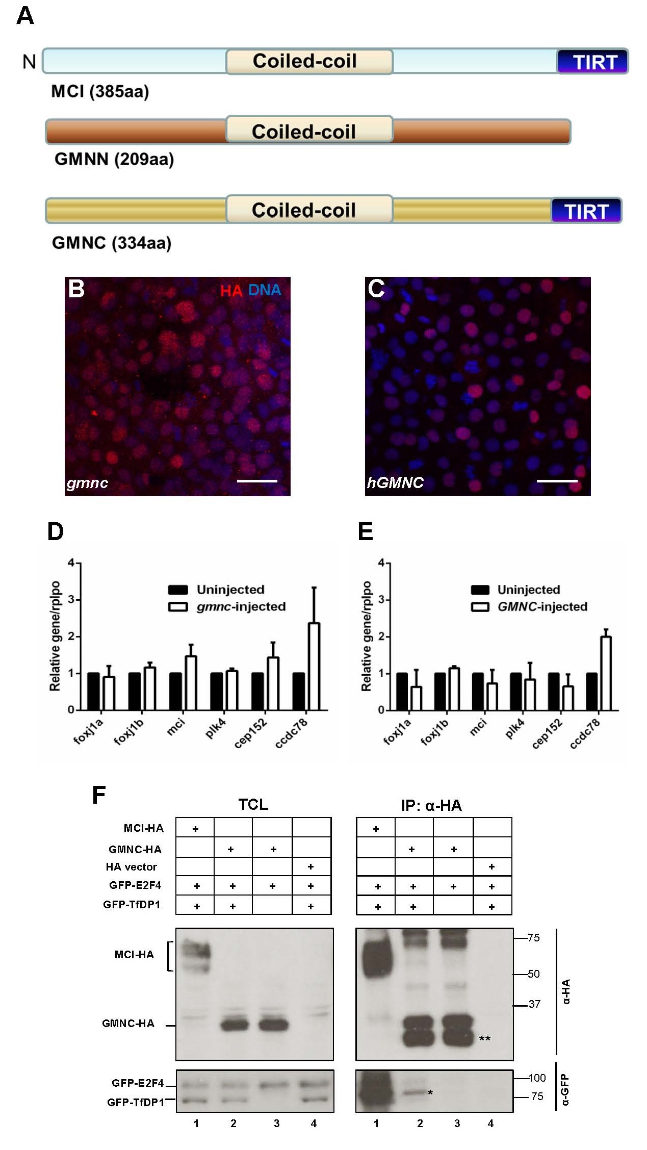
Fig. S3
Schematic of the conserved domains among MCIDAS, GMNN and GMNC, nuclear localization of the zebrafish and human GMNC proteins, the lack of overt transcription activation properties of zebrafish and human GMNC proteins, and the lack of interaction between human GMNC and E2F4/TfDP1
(A) The human MCIDAS, GMNN and GMNC proteins all include a coiled-coil domain. A conserved C-terminus region called the TIRT-domain is present in the MCIDAS and GMNC proteins, but absent in GMNN.
(B and C) In vitro transcribed zebrafish gmnc-HA RNA (encoding Gmnc protein tagged C-terminally with the haemagglutinin (HA) epitope) (B) or human GMNC-HA RNA (C) were injected into one-cell zebrafish embryos. At 7hpf, embryos displayed mosaic expression of the HA-tagged proteins, with nuclear localization. HA (red), DAPI (blue). Scale bars: 10µm.
(D and E) Quantitative qPCRs were performed on zebrafish gmnc-HA (D) or human GMNC-HA (E) RNA-injected embryos at 24 hpf. The ciliogenic genes investigated include foxj1a, foxj1b, mcidas (mci), plk4, cep152 and ccdc78. Expression levels in the uninjected condition were arbitrarily assigned a value of 1. rplpo was used as an internal (loading) control. Error bars represent SEM from 3 (D) or 2 (E) independent experiments.
(F) HA-tagged GMNC (GMNC-HA) was co-transfected in HEK-293T cells with GFP-tagged E2F4 (GFP-E2F4) alone (TCL panel, lane 3) or in the presence of its dimerization partner GFP-TfDP1 (TCL panel, lane 2). Empty HA vector (TCL panel, lane 4) and HAtagged MULTICILIN (MCI-HA) (TCL panel, lane 1) were used as negative and positive controls, respectively. Immunoprecipitation using a monoclonal HA antibody pulled down the MCI-HA and GMNC-HA proteins (IP panel, anti-HA blot). The lower bands (double asterisks) could be degradation products of the GMNC-HA protein. GFP-E2F4/TfDP1 co-immunoprecipitated only with MCI-HA (IP panel, anti-GFP blot, lane 1), but not with GMNC-HA (IP panel, anti-GFP blot, lane 2). The faint band (asterisk) is likely a non-specific species. TCL: Total cell lysate. IP: Immuno-precipitation. The western blot images are representative of the 2 independent experiments performed.