
|
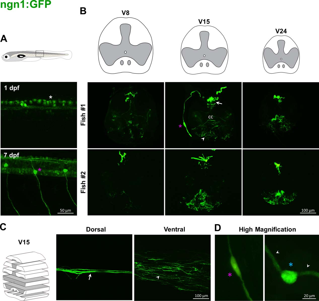
Fig. 7
Labeling pattern of GFP in the spinal cord of adult ngn1:GFP transgenic fish. In ngn1:GFP embryos, the transgene is expressed in Rohon Beard sensory neurons (white asterisk, A, upper panel) and then in neurons of dorsal root ganglia (magenta asterisk, A, lower panel). In adults, GFP-positive fibers lie in dorsal horns (arrows, B, C). A cell body in the dorsal root ganglion and its axon reaching the dorsal horn is stained (magenta asterisks, B; D, left panel). Some processes are labeled at the level of the medioventral white matter (arrowheads, B, C). Higher magnification highlights rare large cells lying in dorsal horn (blue asterisk, D, right panel) with two processes (arrowheads, D, right panel).