
|
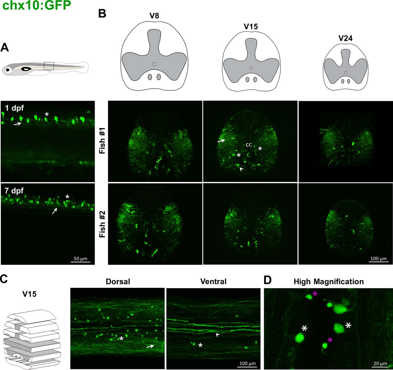
Fig. 5
Labeling pattern of GFP in the spinal cord of adult chx10:GFP transgenic fish. In chx10:GFP embryos, GFP-positive cells are mostly glutamatergic interneurons (asterisk, A) with descending projections (arrow, A). In adults, dense projections are present throughout the gray and white matters (arrows, B, C) except in the more dorsal region. Cell bodies (asterisks, B, C) are located in medial and lateral parts of ventral horns. Some strongly labeled fibers are present in the white matter below the central canal (arrowheads, B, C). At higher magnification large cells stand out from smaller cells (respectively white and magenta asterisks, D).