
Image
|
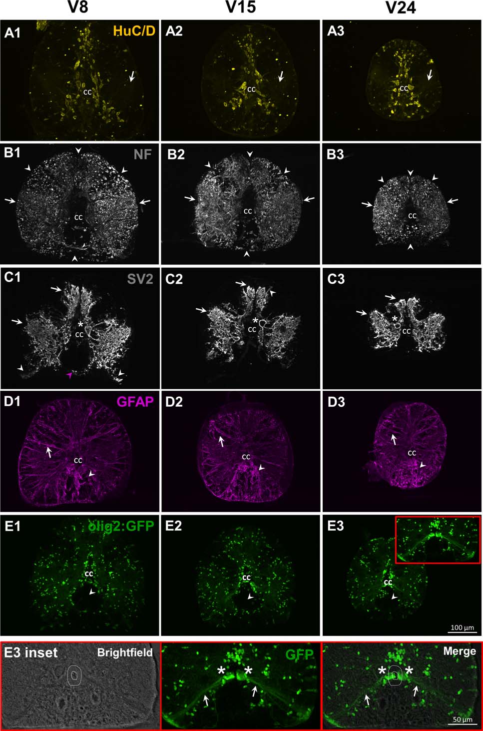
Figure Caption
Fig. 2
Distribution of pan-neuronal and glial markers HuC/D, NF, SV2, GFAP, and Olig2. Neurons are stained with anti-HuC/D antibody (A1-A3). The 3A10 antibody recognizes neurofilament (NF)-associated proteins and labels axonal tracts (B1-B3). The anti-SV2 (synaptic vesicle protein 2) is used to highlight synaptic terminals (C1-C3). Glial cells are identified with the zrf-1 antibody that recognizes glial fibrillary acidic protein (GFAP, D1-D3). Oligodendrocytes are visualized in olig2:GFP transgenic fish (E1-E3).
Acknowledgments
This image is the copyrighted work of the attributed author or publisher, and
ZFIN has permission only to display this image to its users.
Additional permissions should be obtained from the applicable author or publisher of the image.
Full text @ Dev. Neurobiol.