
|
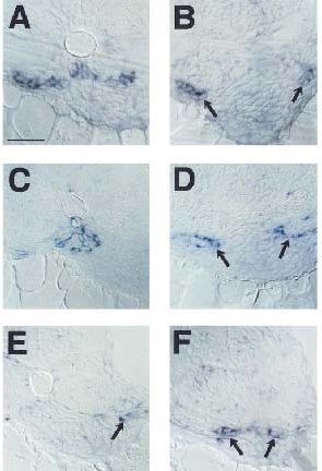
Fig. 3
Glomeruli differentiate at ectopic locations in flh. Wildtype (A, C, E) and flh (B, D, F) embryos hybridized with in situ probes: VEGF (A, B) at 40 hpf, flk-1 (C, D) at 40 hpf, and tie-1 (E, F) at 48 hpf. Differentiated podocytes express VEGF in wildtype embryos (A). VEGF-positive cells are found in ectopic lateral locations in flh (arrows in B). flk-1 labels glomerular capillary-forming endothelial cells in wildtype (C). flk-1-positive cells surround laterally positioned nephric primordia in flh (arrows in D). flk-1-positive cells are found in between nephric primordia and cardinal veins. tie-1 labels endothelia from cardinal veins (arrow in E) but not dorsal aorta in wildtype embryos at this time. In flh, tie-1-positive cells are associated with nephric primordia (arrows in F). Scale bar equals 50 µm.
Reprinted from Developmental Biology, 222(1), Majumdar, A. and Drummond, I.A., The zebrafish floating head mutant demonstrates podocytes play an important role in directing glomerular differentiation, 147-157, Copyright (2000) with permission from Elsevier. Full text @ Dev. Biol.