
|
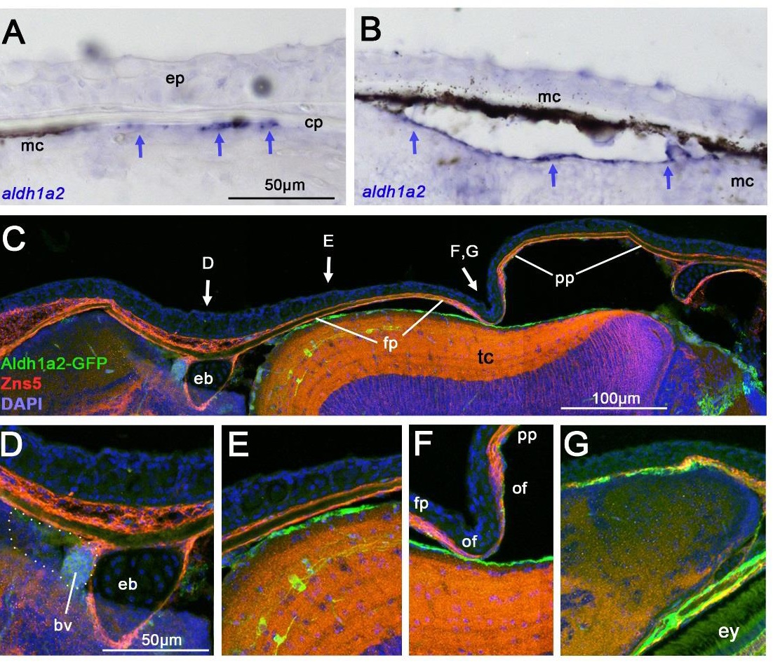
Fig. S3
aldh1a2 displays differential expression in the meninges underlying the calvarial plates. Panels (A,B,G) show transverse sections, panels (C-F) sagittal sections of untreated fish at SL9 (A,B) or SL10-11 (C-G). (A,B) aldh1a2 in situ hybridization; aldh1a2 signals are marked with arrows; melanocytes of the meninges (mc) (Goldgeier et al., 1984) and the calvarial plate (cp) are indicated. Note that the aldh1a2+ cells are positioned between or even underneath the meningeal melanocytes. (C-G) Double immunofluorescence for tg(aldh1a2:aldh1a2-GFP)-driven GFP (Pittlik and Begemann, 2012) and the osteogenic cell marker ZNS5, counterstained with DAPI. Panel (C) shows an overview, and panels (D-F) magnified views of regions indicated in (C); panel (G) shows transverse sections at the level within the region shown in (F). (F,G) are directly at the osteogenic fronts (of) of the frontal (fp) and parietal plates (pp), which have not met and formed a suture as yet; (D) is at the level of the epiphyseal bar (eb), thus, within central regions of the frontal plate and very remote from the osteogenic front, and (E) is in an intermediate position closer to the frontal pate osteogenic front. In (D), the distinct and highly vascularized tissue directly anterior of the epiphyseal bar, which displays strong RA-induced cyp26b1 expression (Fig. 7I-L), is outlined. The Aldh1a2-GFP signal is much stronger in regions close to the osteogenic front than in very remote regions. In addition, it is not localized within the ZNS5-positive osteogenic cells themselves, but in cells below them, at about the same level like the meningeal melanocytes. This expression pattern correlates with the pattern of calvarial growth, with horizontal growth occurring at the osteogenic fronts (see Fig. 1D), and vertical growth preferentially occurring on the inner surface of the calvarial plates (see Fig. 1G). Abbreviations: bv, blood vessel; cp, calvarial plate; eb, epiphyseal bar; ep, epidermis; ey, eye: fp, frontal plate; mc, melanocytes; of, osteogenic front; pp, parietal plate, tc, optic tectum.