
|
Fig. 1
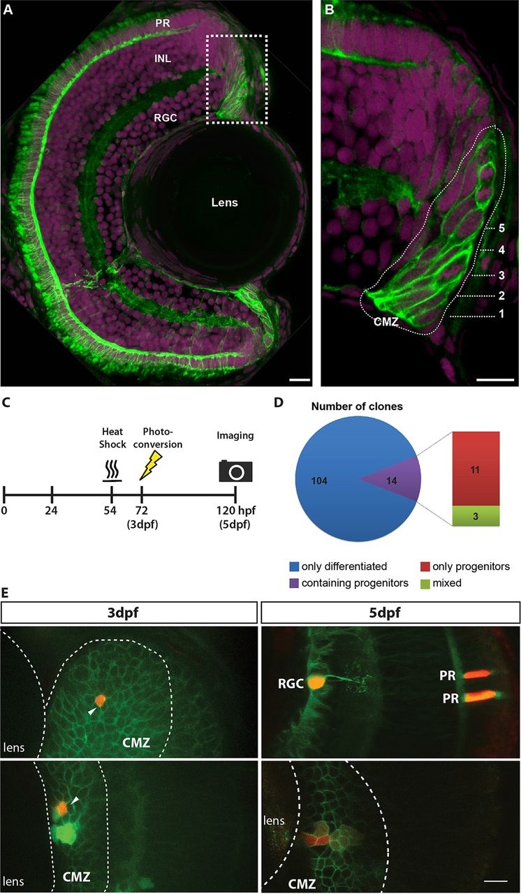
Short-term clonal analysis. Rx2 functions as a marker for the CMZ of the zebrafish retina. (A) A maximum intensity projection of a transverse 12µm section of a 5dpf zebrafish retina, showing the expression of the transgenic reporter Tg(Rx2:GFPcaax). Rx2+ cells (green) are found in the CMZ as well as in two cell types of the neural retina, photoreceptors (PRs) and Müller glia. (B) High-magnification 1µm section from a region of the CMZ (box in A), revealing that both RSCs (rings 1-2) and RPCs (rings 3-5) are Rx2+. Numbers indicate rings. Nuclei are labelled with DAPI (magenta). INL, inner nuclear layer; RGC, retinal ganglion cell layer. (C) Experimental procedure for labelling CMZ cells and capturing the short-term clonal composition. (D) Composition of short-term CMZ clones reveals the distinct proliferative fates in the CMZ population. (E) A single CMZ cell is photoconverted to red at 3dpf (top left, indicated by arrow) and the resultant terminated clone at 5dpf (top right, red) is shown with identified cell fates (RGC, retinal ganglion cell; PR, photoreceptors). Another example of a single CMZ cell photoconverted at 3dpf (bottom left, indicated by arrow) and the resultant maintained clone observed at 5dpf (bottom right, red). Note the dilution of the label in cells further from the lens, which is suggestive of more rapid division. In E, all Rx2+ cells have green membranes whereas the MAZe:Kaede cells have green cytoplasm that becomes red upon photoconversion. Scale bars: 10µm.