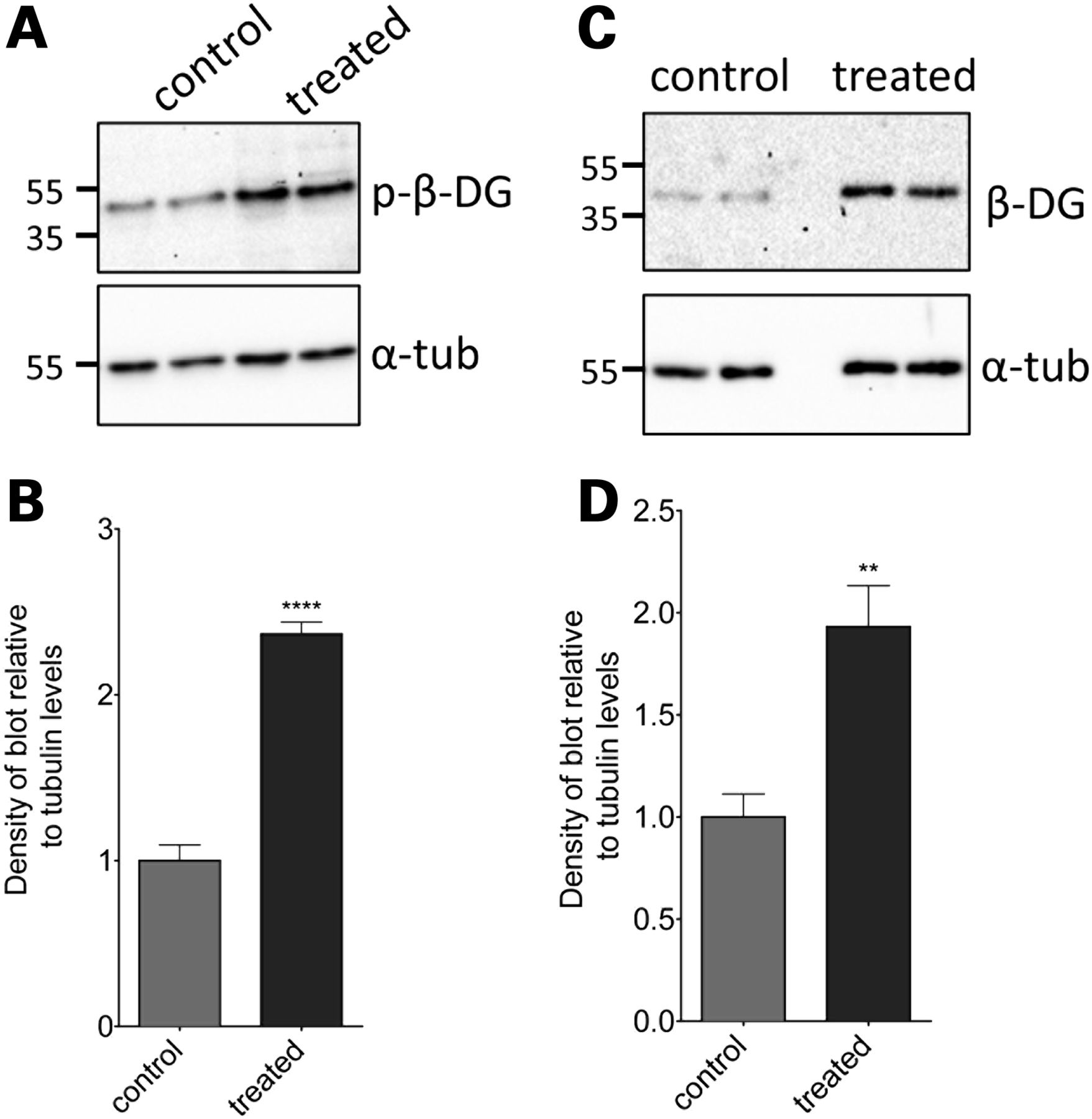
Fig. 6 The effect of MG132 treatment on levels of phosphorylated and non-phosphorylated β-dystroglycan in sapje zebrafish larvae. Lysates were made from sapje/ embryos treated from 24 hpf until 96 hpf with 5 μM MG132 or DMSO-only. (A) Western blots probed with antibodies for β-dystroglycan (β-DG) and α-tubulin (α-tub). (B) The density of the blot probed with β-DG was quantified relative to α-tubulin levels in each sample, and normalized to the average control signal. There was a significant increase in the level of β-dystroglycan in larvae treated with MG132, compared with controls (unpaired t-test: t = 4.048, df = 10, P = 0.0023). (C) Western blots probed with antibodies for phosphorylated β-dystroglycan (p-β-DG) and α-tubulin. (D) The density of the blot probed with p-β-DG was quantified relative to α-tubulin levels in each sample, and normalized to the average control signal. There was a significant increase in the levels of β-DG in larvae between different treatment groups (unpaired t-test: t = 11.47, df = 10, P < 0.0001). Graphs represent the mean of six samples from three independent experiments, error bars are SEM.
Image
Figure Caption
Figure Data
Acknowledgments
This image is the copyrighted work of the attributed author or publisher, and
ZFIN has permission only to display this image to its users.
Additional permissions should be obtained from the applicable author or publisher of the image.
Full text @ Hum. Mol. Genet.

