
|
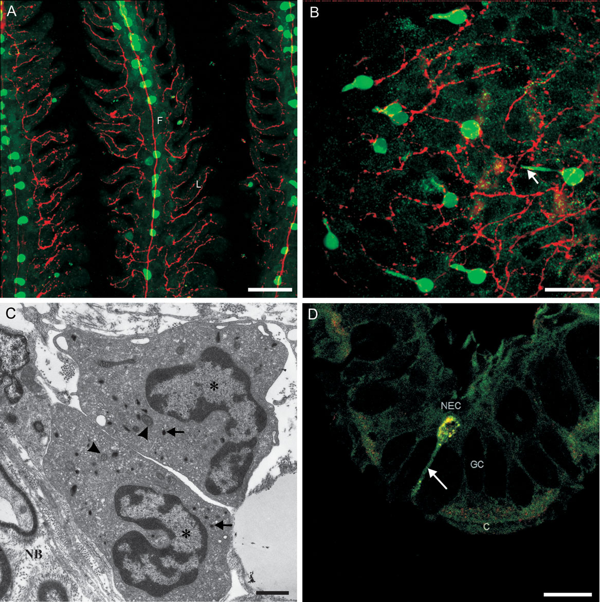
Fig. 1
Neuroepithelial cells (NECs) in gills, skin, and respiratory gas bladder in fish are neurosecretory and contain serotonin (5-HT). (A) NECs of gill filaments (F) and lamellae (L) of adult zebrafish were labeled with antibodies against 5-HT (green), and innervating nerve fibers were labeled with a neuron-specific marker (zn-12, red). Scale bar = 50 µm. Confocal microscopy and immunolabeling procedures were similar to those described in Ref. 22. (B) NECs and associated nerve fibers of the yolk sac epithelium of a 7-dpf zebrafish larva were labeled in the same manner as in A. Note the long processes (arrow) that extend to make contact with the external environment. Scale bar = 20 µm. (C) Transmission electron micrograph of two NECs from Lepisosteus oculatus respiratory bladder showing heterochromatin nucleus (asterisks), small mitochondria (arrowheads), numerous profiles of smooth and rough endoplasmic reticulum, and specific, membrane-bound, irregularly shaped vesicles with dense content (arrows). Note their close relationship with nerve bundle (NB)-containing myelinated axons. Scale bar = 1 µm. Modified with permission from Ref. 48. (D) Immunocytochemical double staining for calbindin D28k (red) and 5-HT (green) of a NEC in the mucociliated epithelium of the respiratory gas bladder of L. oculatus. The confocal image shows a combination of the two color channels and the orientation of the cell making contact with the external surface by a slender process (arrow). GC, goblet cell; C, ciliated cell. Scale bar = 10 µm.