
|
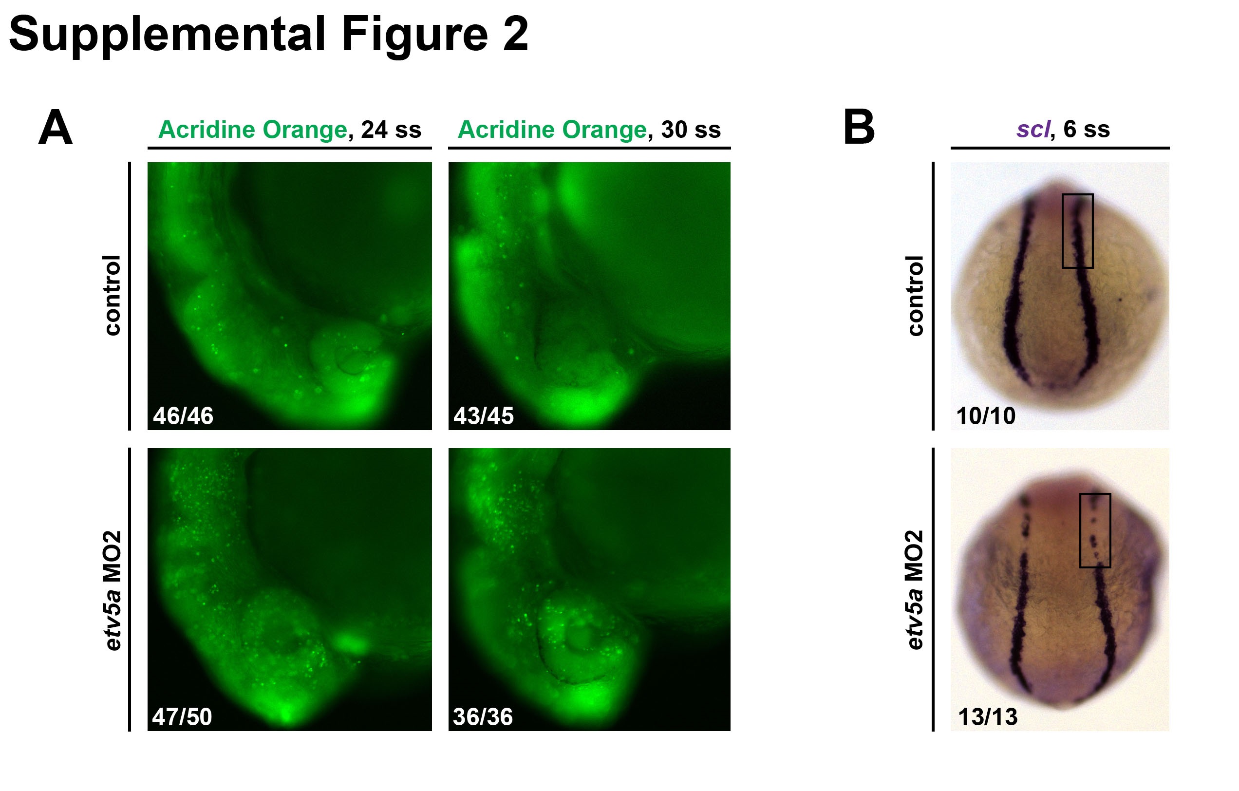
Fig. S2 etv5aMO2 assays. (A) Embryos injected with etv5a MO2 have more cell death, as shown by AO staining, in the head region compared to control embryos at the same time points. However, the amount of cell death is reduced in etv5a MO2 injected embryos from the 24 ss to the 30 ss. (B) WISH analysis on 6 ss embryos shows reduced scl (purple) expression in embryos injected with etv5a MO2. Embryo counts for each observed phenotype are as indicated in the lower left corner of the corresponding panel. For control 30 ss, 2/45 embryos had elevated AO cell numbers compared to the majority phenotype that is shown, and for etv5a MO2 24 ss, 3/50 embryos had further elevated AO cell numbers compared to the majority phenotype that is shown.
Reprinted from Developmental Biology, 411(2), Marra, A.N., Wingert, R.A., Epithelial cell fate in the nephron tubule is mediated by the ETS transcription factors etv5a and etv4 during zebrafish kidney development, 231-45, Copyright (2016) with permission from Elsevier. Full text @ Dev. Biol.