
|
Fig. 5
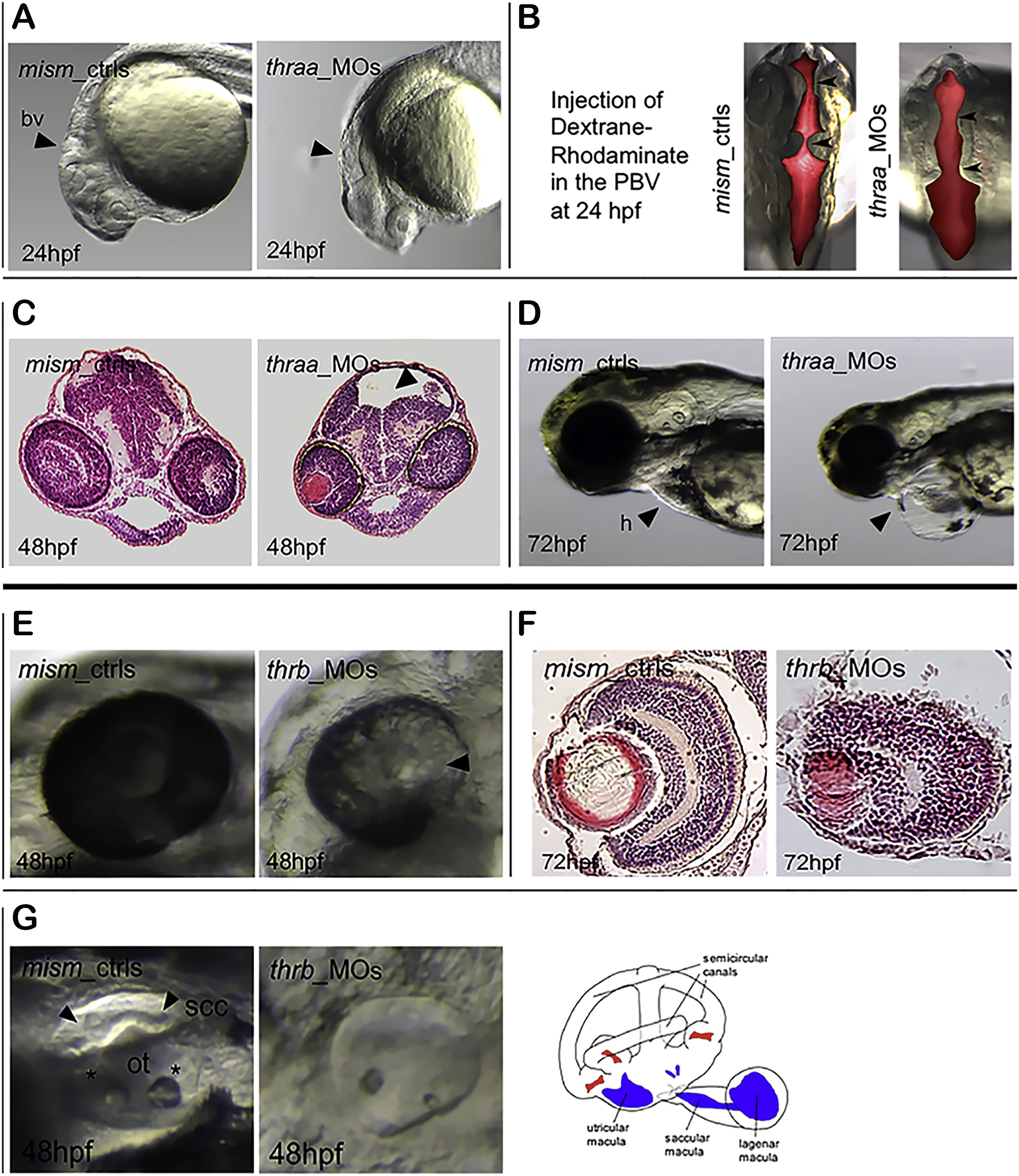
Panels A-D) Morphological defects of thraa _MOs. At 24 hpf, thraa_MOs display small head (Panel A, arrow) and a dilatation of brain ventricles (bv, arrows) with an accumulation of dextrane-rhodaminate dye (dorsal view, Panel B). At 48 hpf, histological sections confirm the hydrocephalus in thraa_MOs (Panel C, arrow). At 72 hpf, the heart (h) edema is visible in thraa_MOs (Panel D, arrow). Panels E-G) Morphological defects of thrb_MOs. At 48 hpf, the thrb_MOs present hypopigmented eyes (Panel E) with a defective development of retina visible in the histological sections at 72 hpf (Panel F). Alterations of the otic vesicle structures in thrb_MOs at 48 hpf (Panel G): ssc, semicircular canals (arrows); ot, otoliths (asterisks). The schematic structure of the otic vesicle is illustrated in the drawing image from Whitfield TT (Whitfield, 2015).
Reprinted from Molecular and Cellular Endocrinology, 424, Marelli, F., Carra, S., Agostini, M., Cotelli, F., Peeters, R., Chatterjee, K., Persani, L., Patterns of thyroid hormone receptor expression in zebrafish and generation of a novel model of resistance to thyroid hormone action, 102-17, Copyright (2016) with permission from Elsevier. Full text @ Mol. Cell. Endocrinol.