
|
Fig. 3
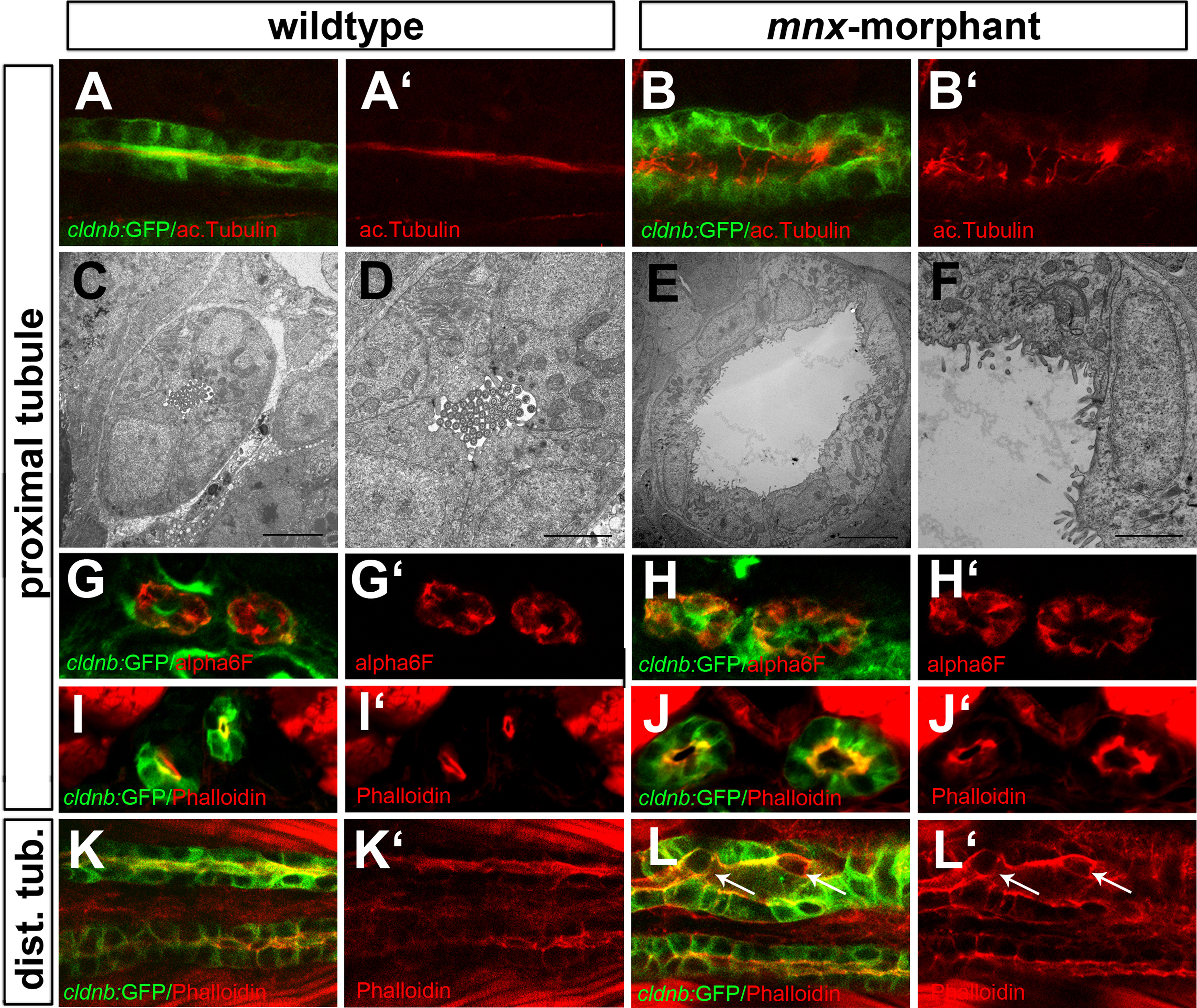
Mnx knockdown causes defects in tubular cilia arrangement and epithelial morphology. (A, B) Single plane confocal images showing tubule cilia stained by anti-actelytaled tubulin (red). Shown are lateral views of proximal tubules of control (A, A′) and mnx-morpholino injected Tg(cldnb:lynGFP) embryos (B, B) (C-F) Transverse TEM sections of proximal tubules in control (C, D) and mnx-morpholino embryos (E, F). Note disorganized microvilli structures after mnx gene knockdown (scale bars correspond to 5 µm (C, E) and 2 µm (D, F)). (G-J) Transverse sections of proximal tubules in control and mnx morphant embryos at 2dpf. Staining of baso-lateral surface directed alpha6F (G, H) and apically located Phalloidin (I, J) displayed similar expression patterns in control and MO-mnx. (K, L) Single plane confocal images of distal tubules stained for Phalloidin (red, ventral view). White arrows indicate displaced epithelial cells with unpolarized Phalloidin signals.
Reprinted from Developmental Biology, 411(1), Ott, E., Wendik, B., Srivastava, M., Pacho, F., Töchterle, S., Salvenmoser, W., Meyer, D., Pronephric tubule morphogenesis in zebrafish depends on Mnx mediated repression of irx1b within the intermediate mesoderm, 101-14, Copyright (2016) with permission from Elsevier. Full text @ Dev. Biol.