
Image
|
Figure Caption
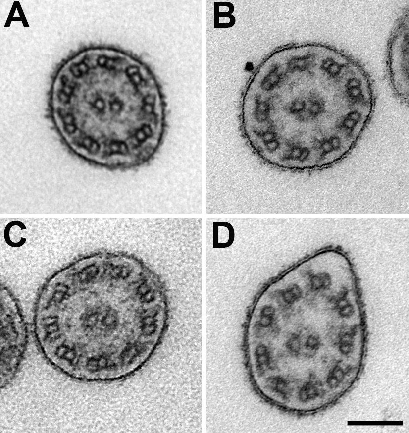
Fig. S6
Normal cilia structure in mnx2b and irx1b morphants. TEM images of cilia from the posterior part of the pronephric duct of 3dpf embryos showing regular ultrastructure. A: control, B: irx1b-MO, C: mnx2b-MO 1:2, D: mnrx2b-MO + irx1b-MO. Scale bar is 100 nm
Acknowledgments
This image is the copyrighted work of the attributed author or publisher, and
ZFIN has permission only to display this image to its users.
Additional permissions should be obtained from the applicable author or publisher of the image.
Reprinted from Developmental Biology, 411(1), Ott, E., Wendik, B., Srivastava, M., Pacho, F., Töchterle, S., Salvenmoser, W., Meyer, D., Pronephric tubule morphogenesis in zebrafish depends on Mnx mediated repression of irx1b within the intermediate mesoderm, 101-14, Copyright (2016) with permission from Elsevier. Full text @ Dev. Biol.