
|
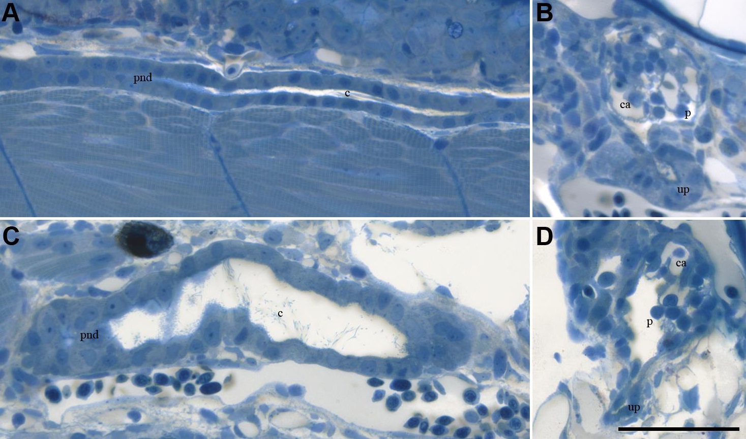
Fig. S4
Histology of tubule and glomeruli in 3 dpf mnx2b morphants. Histological horizontal section of a pronephric duct (A, B) and of a glomerolus (B, D) in control animal (A, B) and in mnx2b morphant (C, D). Dilated pronephric duct (C) and glomerolus of a mnx2b morphant (D). Note the urinary pole on both glomerula (up). ca = capillary, c = cilia, p = podocyte, pnd = pronephric duct. Scale bar is 50 µm. Rombout fixed and Polybed 812 embedded zebrafish were cut serially with a Histo Butler diamond knife (Diatome, Switzerland) with a thickness of 2 µm, stained with 1% methylen blue and 1% AZUR II in 1% sodium tetraborate for 2 minutes and examined with a leica DM5000B microscope. Images were made with a Leica DFC490 digital camera and aLeica application suite V4 software.
Reprinted from Developmental Biology, 411(1), Ott, E., Wendik, B., Srivastava, M., Pacho, F., Töchterle, S., Salvenmoser, W., Meyer, D., Pronephric tubule morphogenesis in zebrafish depends on Mnx mediated repression of irx1b within the intermediate mesoderm, 101-14, Copyright (2016) with permission from Elsevier. Full text @ Dev. Biol.