
Fig. 2
- ID
- ZDB-IMAGE-160324-17
- Publication
- Chang et al., 2016 - CDX4 and retinoic acid interact to position the hindbrain-spinal cord transition
- All Figures
- Figures for Chang et al., 2016
|
Fig. 2
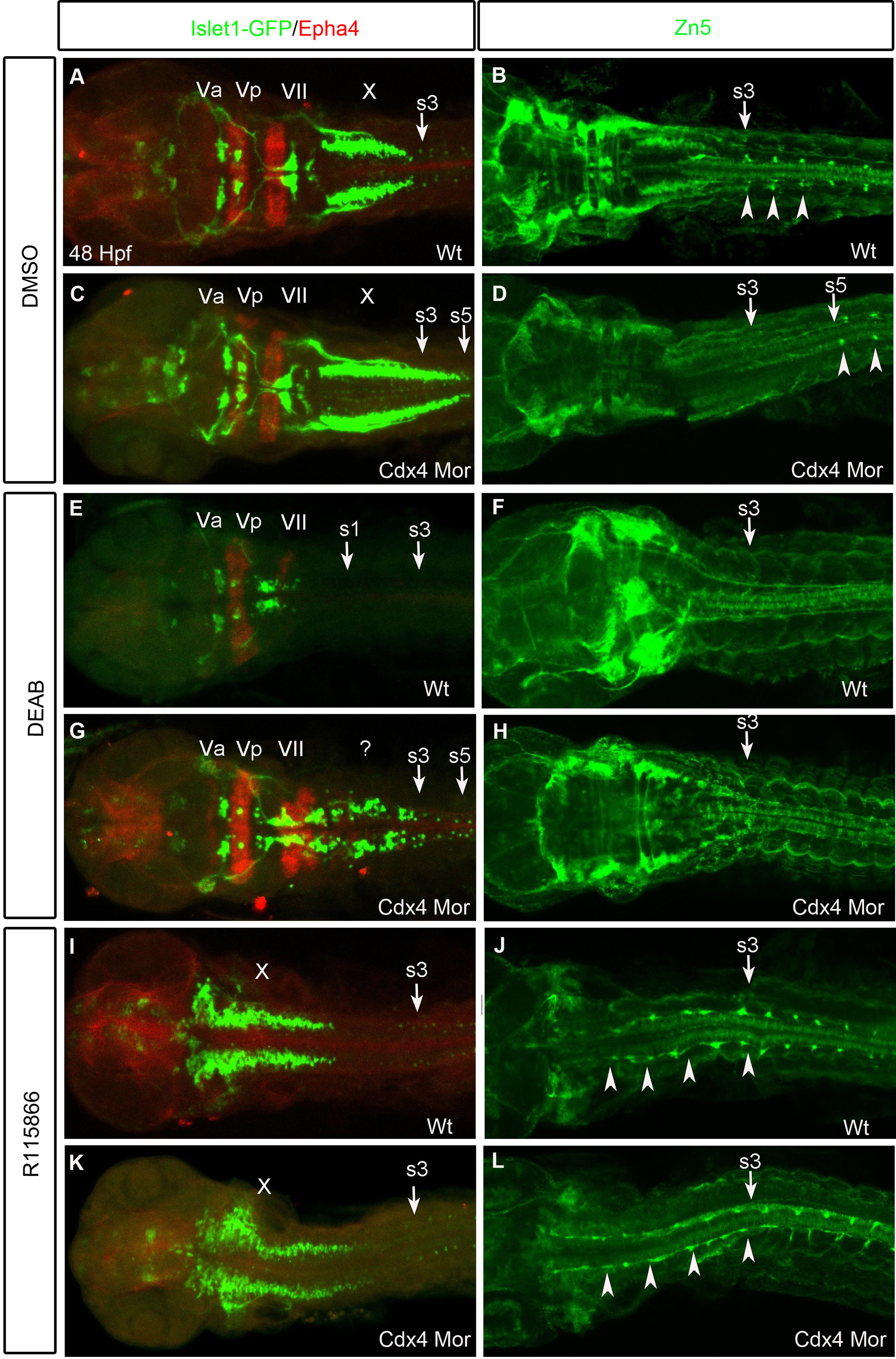
Hindbrain and spinal cord neural populations shift along the AP-axis when Cdx4 or RA functions are perturbed. (A,C,E,G,I,K) Branchial motor neurons marked by Islet1-Gfp and r3,r5 visualized by anti-Epha4. The trigeminals(Va/Vp) in r2-3 and facials(VII) in r6 are unchanged but the vagals(X) in r7/8 are caudally expanded in Cdx4-deficient embryos (C) compared to wildtypes (A). Vagals (r7/8) and Epha4 (r5) are lost in RA-deficient embryos (E). Epha4 in r5 and Islet1-Gfp cells in r5-8 are rescued in RA/Cdx4 deficient embryos (G). Vagals are rostrally shifted while trigeminals and facials are absent in both wildtypes (I) and Cdx4-deficient embryos (K) treated with R115866. (B,D,F,H,J,L) Spinal cord motor neurons marked by Zn5 antibody. The first spinal cord motor neuron arises at the level of somite 3 in wildtypes (B arrow) but at the level of somite 5 in Cdx4-deficient embryos (D arrow). Spinal cord motor neurons are lost in RA-deficient (F) and RA/Cdx4 deficient (H) embryos. Spinal cord motor neurons expand rostrally in both wildtypes (J) and Cdx4-deficient embryos (L) treated with R115866. (A-L) Flat-mounted embryos at 48 hpf. (A,C,E,G,I,K) n=30/30 per condition and (B,D,F,H,J,L) n=15/15 per condition.
Reprinted from Developmental Biology, 410(2), Chang, J., Skromne, I., Ho, R.K., CDX4 and retinoic acid interact to position the hindbrain-spinal cord transition, 178-89, Copyright (2016) with permission from Elsevier. Full text @ Dev. Biol.