
Fig. 1
- ID
- ZDB-IMAGE-160324-16
- Publication
- Chang et al., 2016 - CDX4 and retinoic acid interact to position the hindbrain-spinal cord transition
- All Figures
- Figures for Chang et al., 2016
|
Fig. 1
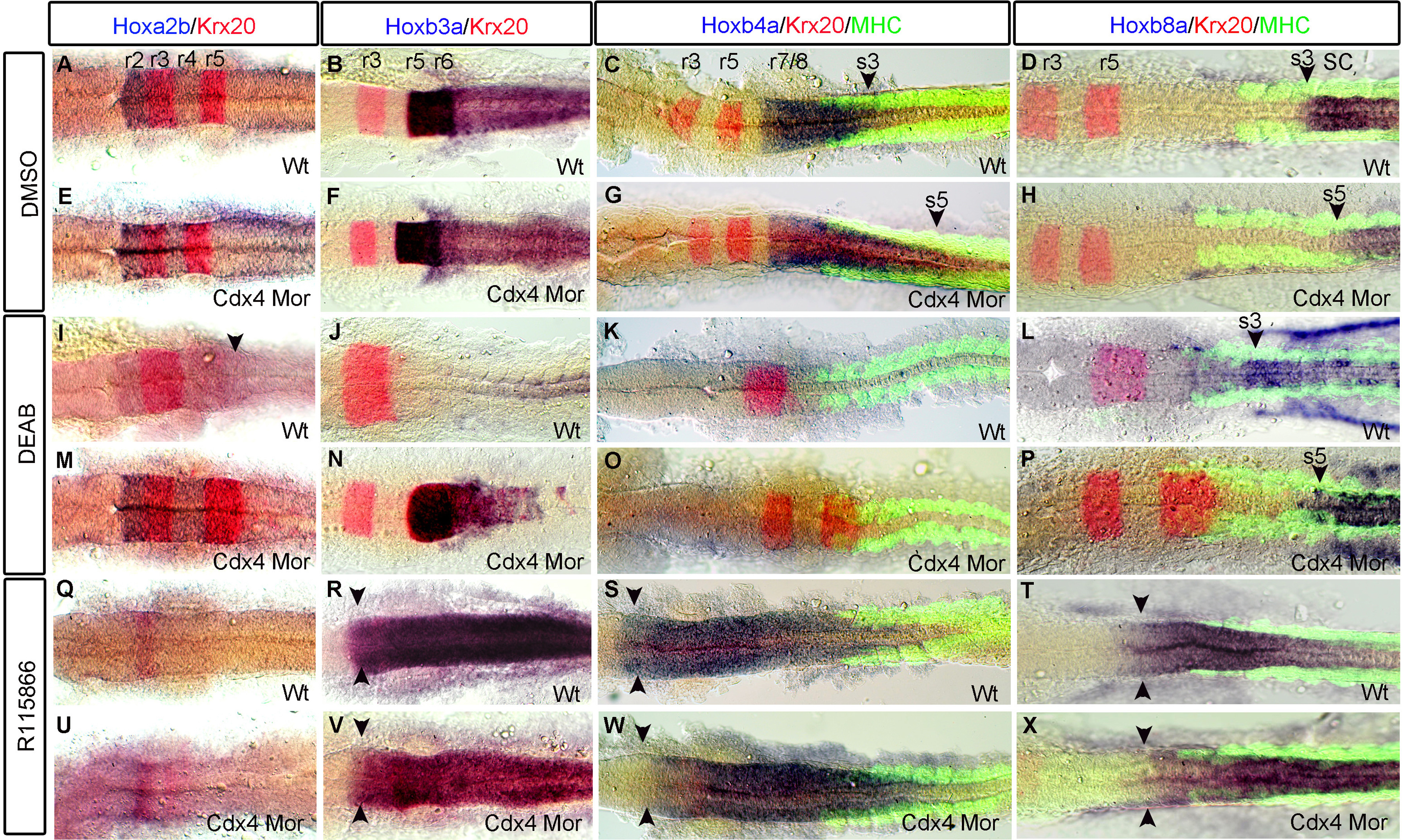
Hindbrain and spinal cord hox gene expression shifts along the AP-axis when Cdx4 or RA functions are perturbed. Expression of hoxa2b in r2-4 and hoxb3a in r5-6 is the same in Cdx4-deficient embryos (E, F) and wild-types (A, B). hoxb4a expression in r7/8 is caudally expanded in Cdx4-deficient embryos (G arrowhead) compared to wildtypes (C arrowhead). In RA-deficient embryos hoxb3a (J) and hoxb4a (K) expression is lost, while hoxa2b expression in r4 is caudally expanded (I arrowhead). In RA/Cdx4 deficient embryos, hoxb3a expression is rescued (N) but not hoxb4a (O). In both wild type and Cdx4 deficient embryos treated with R115866 hoxa2b (Q, U) expression is lost while hoxb3a (R, V) and hoxb4a (S, W) expression are both rostrally expanded. The anterior expression limit of spinal cord hoxb8a is located at somite 3 in wildtypes (D arrowhead) and RA deficient embryos (L arrowhead) but its expression is caudally shifted to somite 5 in Cdx4 deficient embryos (H arrowhead) and RA/Cdx4 deficient embryos (P arrowhead). hoxb8a expression expands into the hindbrain in both wild type (T arrowheads) and Cdx4 deficient embryos (X arrowhead) treated with R115866. (A-X) are co-stained with krx20 (r3,r5). (C, D, G, H, K, L, O, P, S, T, W, X) co-stained with myosin heavy chain antibody (somites). (A-X) flat-mounted embryos at 20 somite stage n=45/45 per condition.
Reprinted from Developmental Biology, 410(2), Chang, J., Skromne, I., Ho, R.K., CDX4 and retinoic acid interact to position the hindbrain-spinal cord transition, 178-89, Copyright (2016) with permission from Elsevier. Full text @ Dev. Biol.