
|
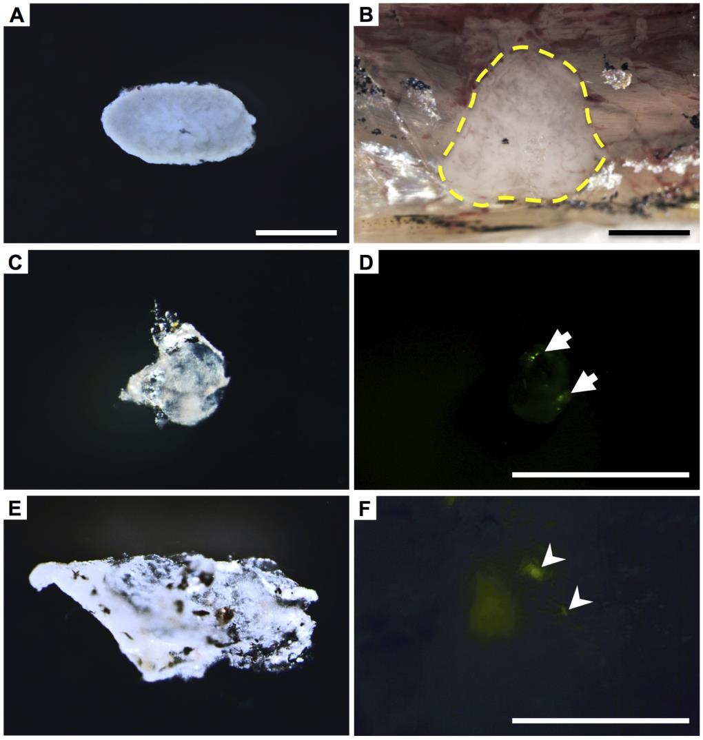
Fig. S1
Growth of normal testicular fragments and testicular aggregates mixed with SSCs of testicular hyperplasia after transplantation. (A, B) The normal testicular fragment prior to the transplantation into rag1t26683 mutant hosts (A), and the grafted testis fragment (B, enclosed by a dotted line) uncovered by peeling of host’s skin after 2 months of transplantation. (C, D) The aggregate of the inbred IM line that was mixed with spermatogonia of normal vas::egfp testes after 3 months of transplantion in the IM line. Arrows indicate GFP-positive cells (D). (E, F) The aggregate of the inbred IM line that was mixed with cultured SSCs of vas::egfp hyperplasias after 3 months of transplantation in the IM line. The SSCs were cultured for 4 weeks in the previous culture condition (Kawasaki et al., 2012). Arrowheads indicate GFP-positive cells (F). Sperm obtained from this aggregate generated fertilized embryos. Scale bar, 1mm.