
|
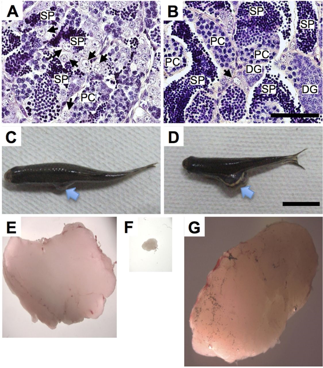
Fig. 1
Maintenance and growth of testicular hyperplasias following subcutaneous transplantation. (A,B) Histological observation of a testicular hyperplasia (A) and normal testis (B). Arrows indicate a single spermatogonium surrounded by Sertoli cells. DG, differentiating spermatogonia in large clusters surrounded by Sertoli cells; PC, primary spermatocytes; SP, sperm. Note that the hyperplasia contains a large number of single spermatogonia. Scale bar: 50µm. (C-G) Views of the rag1t26683 mutant just after transplantation beneath the abdominal skin (C) and after 3months (D). A testicular hyperplasia (E) was cut down to transplantable size (F). After 3 months of transplantation, the grafted fragment was removed (G). Note that the grafted region swelled (arrows in C,D) and the transplanted fragment grew to almost original size (E,G). Scale bar: 1cm in C,D and 3.3mm in E-G.