
|
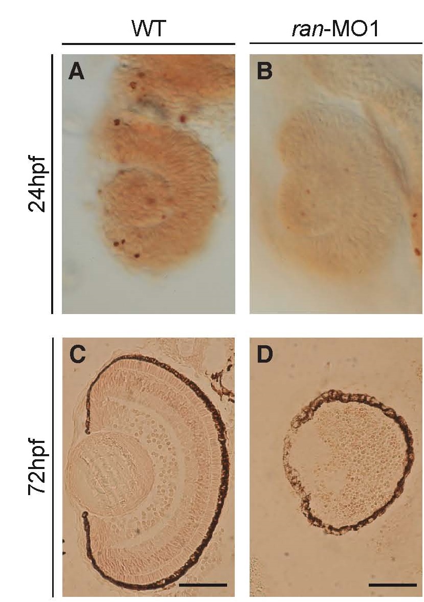
Fig. S3
Ran-deficient embryos did not exhibit a significant change in the apoptosis of retinal cells. (A,B) Lateral views of 24 hpf eyes. (C,D) coronal sections of 72 hpf eyes. The apoptosis signals in the embryonic ocular section were detected by TUNEL assay. A small amount of apoptosis signals was detected in both wild-type embryos and ran-deficient embryos at 24 hpf, in which there was not much differences. Additionally, few apoptosis signals were also detected in both wild-type embryos and ran-morphants at 72 hpf, indicating that apoptosis would not be caused by the loss of Ran at both early and late developmental stages of retinal cells. Scale bars, 50 µm.