
Image
|
Figure Caption
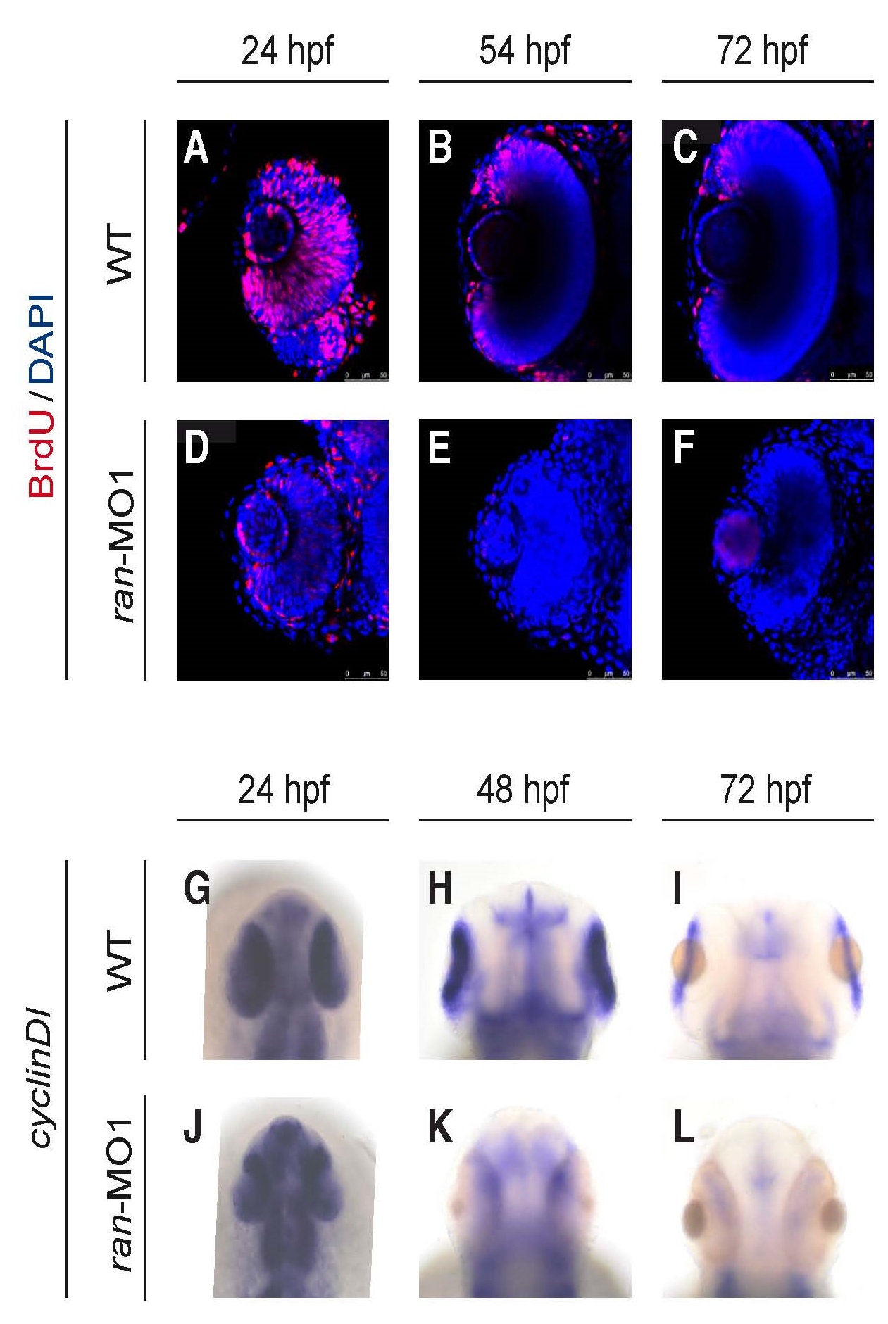
Fig. 4
Ran deficiency impaired eye cell proliferation. (A-F) Temporal dynamics of cell proliferation on the eyes of WT and ran-deficient embryos. All images are coronal sections with anterior to the top. Red, BrdU; Blue, DAPI. Scale bars indicate 50 µm. (G-L) Temporal dynamics of cell proliferation marker cyclinD1 mRNA on the eyes of WT and ran-deficient embryos. All images are dorsal view with anterior to the top. All images are representative of at least three repeats with n>25 for each repeat.
Figure Data
Acknowledgments
This image is the copyrighted work of the attributed author or publisher, and
ZFIN has permission only to display this image to its users.
Additional permissions should be obtained from the applicable author or publisher of the image.
Full text @ Int. J. Dev. Biol.