
|
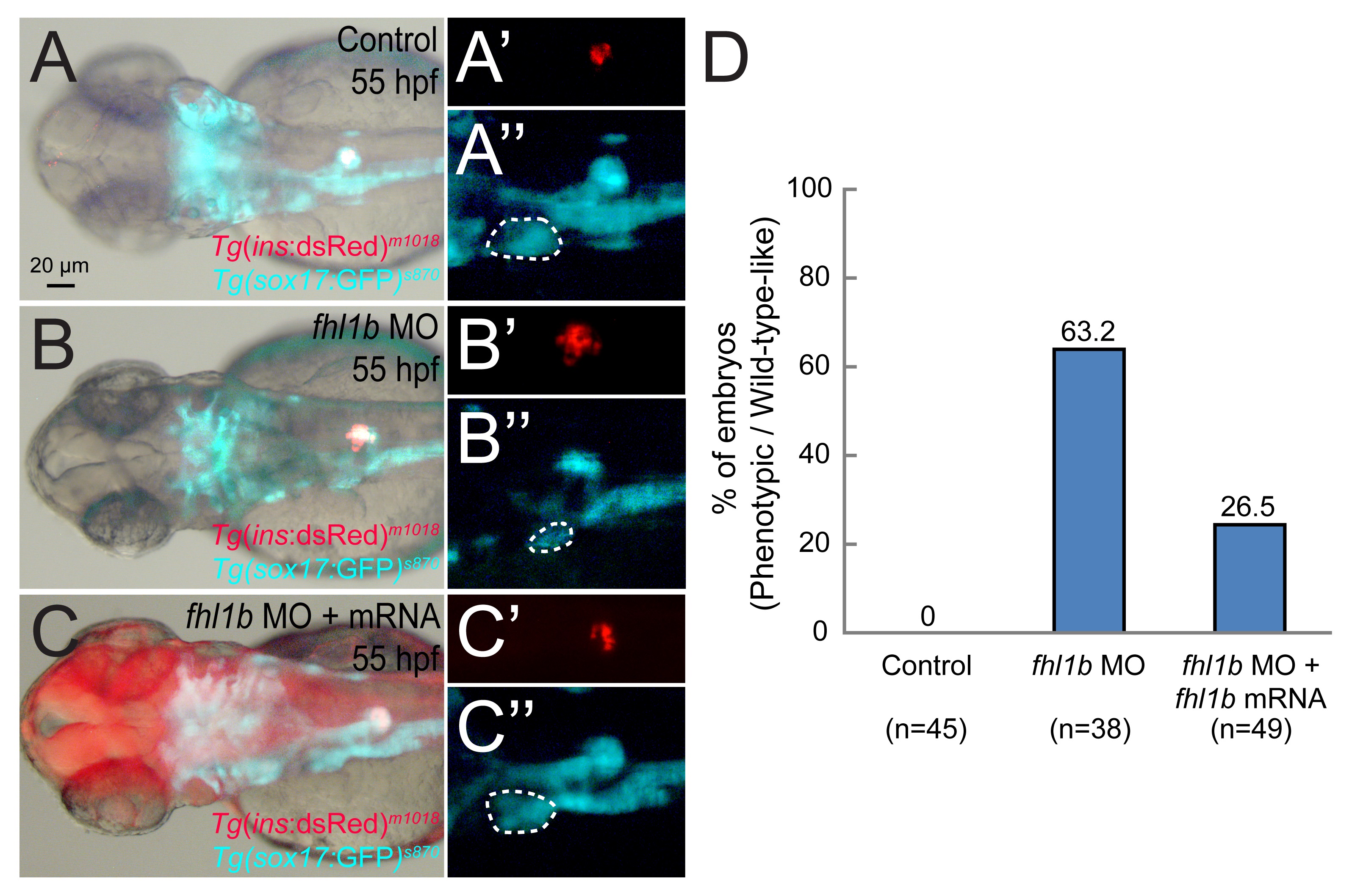
Fig. S6
fhl1b mRNA injection partially rescues the effect of fhl1b MO knockdown.
(A-C′′) The developmental defects of the liver (A-C and white dotted circles in A′′-C′′) and β-cell formation (A-C and A′-C′) in fhl1b morphants (B-B′′) could be partially rescued by injection of fhl1b-P2A-mcherry mRNA (C-C′′), restoring liver size and the β-cell population to a degree comparable to that of control embryos (A-A′′) at 55 hpf. Fhl1b translation was monitored by mCherry expression as shown in C. (D) Quantification of the results in A-C′′. The embryos were scored as having a “reduced” or “increased” expression domain when the expression area of Tg(ins:dsRed)m1018 and Tg(sox17:GFP)s870 was distinctly (> 25%) smaller or larger than that of the control embryos based upon the calculation using ImageJ. A-C, bright-field images combined with fluorescent image of Tg(ins:dsRed)m1018 and Tg(sox17:GFP)s870 expression. A′-C′′, fluorescent images of Tg(ins:dsRed)m1018 and Tg(sox17:GFP)s870 expression. Dorsal views, anterior to the left. Scale bar, 20 µm.