
|
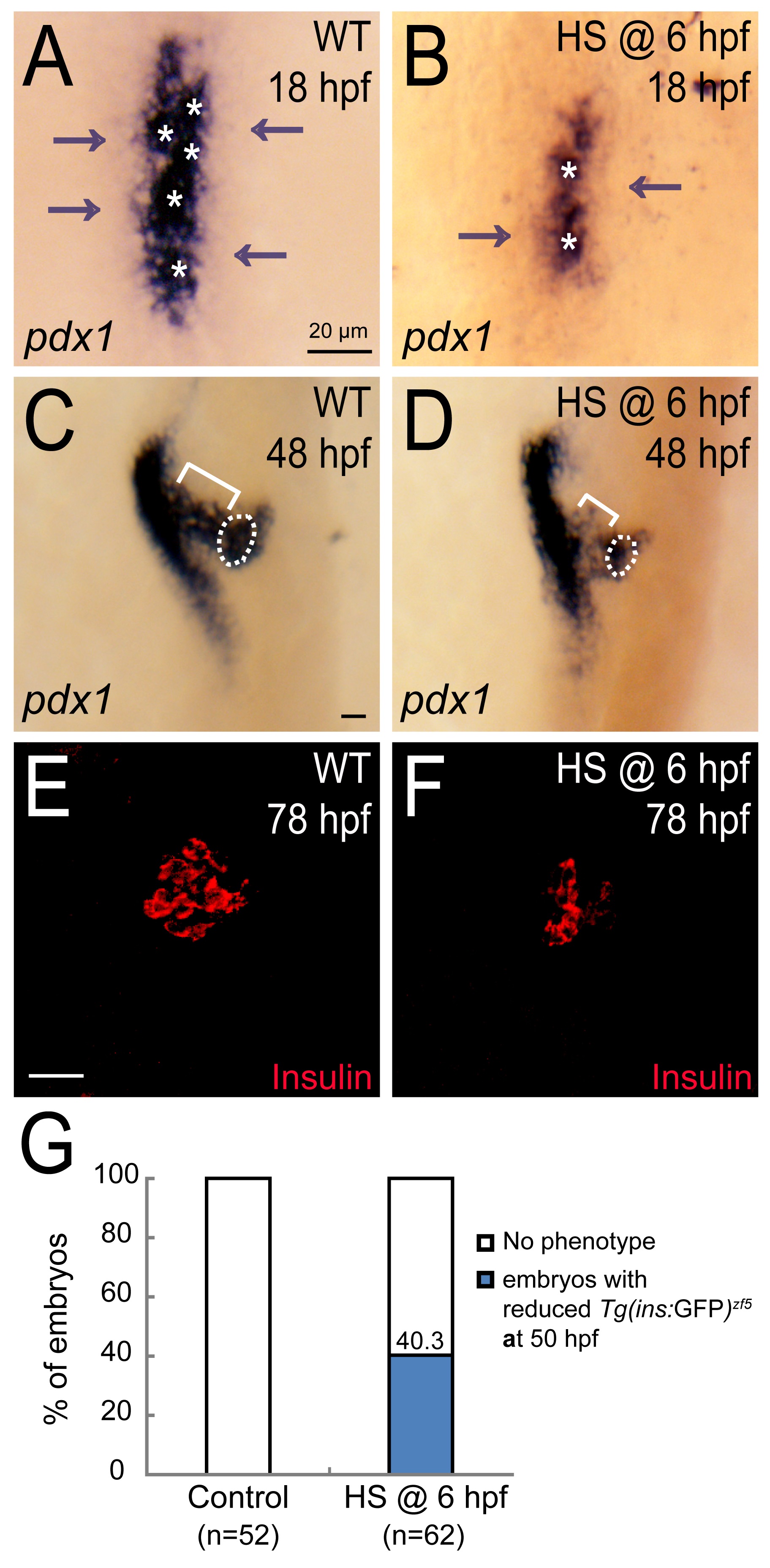
Fig. S12
Forced induction of fhl1b during the gastrulation stage directly or indirectly modulates pdx1 levels for the endocrine and exocrine pancreas development.
(A-D) Whole-mount in situ hybridization showing the expression of pdx1 at 18 hpf (A-B) and 48 hpf (C-D), comparing control embryos (A and C) and fhl1b-overexpressing embryos (B and D, heat shock applied at 6 hpf). In embryos induced to overexpress fhl1b at 6 hpf, both high (white asterisks) and low (gray arrows) levels of pdx1 expression were reduced at 18 hpf (B). Consistently, at 48 hpf, pdx1 expression in the principal islet (white dotted circle) and in the developing exocrine pancreas (white bracket) was reduced (D). (E and F) Confocal images of control embryos and embryos induced to overexpress fhl1b at 6 hpf, stained for Insulin (red). The number of insulin cells was reduced in fhl1b-overexpressing embryos (F), compared to that of control embryos (E) at 78 hpf. (G) The expression of Tg(ins:GFP)zf5 (heat shock applied at 6 hpf) was examined at 50 hpf and the percentages of embryos were quantified. The embryos were scored as having a “reduced” expression when the expression of Tg(ins:GFP)zf5 was distinctly (> 25%) smaller than that of the control embryos based upon the calculation using ImageJ. A-D, dorsal views, anterior to the top. E-F, confocal projection images, ventral views, anterior to the top. Scale bars, 20 µm.