
|
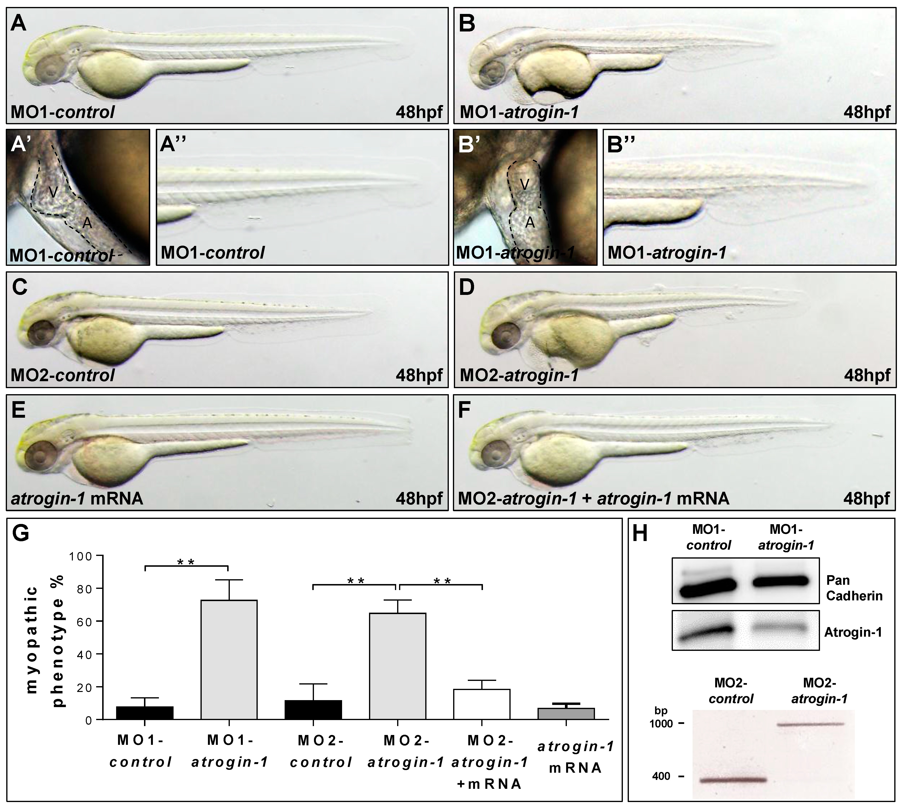
Fig. 2
Knock-down of Atrogin-1 leads to defects in muscle and heart morphology. (A,B) Lateral view of embryos at 48 hpf injected either by 5 bp mismatch Start Morpholino (MO1-control, A) or Start Morpholino directed against atrogin-1 (MO1-atrogin-1, B); (A′,A′′-B′,B′′) Close-ups of heart and tail of injected embryos at 48 hpf; (C,D) Lateral view of embryos at 48 hpf either injected by a 5 bp mismatch Morpholino targeting a splice donor site of Atrogin-1 as a control (MO2-control) or a Splice Morpholino (MO2-atrogin-1); (E,F) Lateral view of embryos at 48 hpf. Control injection with atrogin-1 mRNA (E); Rescue of the morphant phenotype by injecting atrogin-1 mRNA and MO2-atrogin-1 together (F); (G) Statistical analysis of affected embryos after MO1/2-control, MO1/2-atrogin-1, atrogin-1 mRNA or MO2-atrogin-1 and atrogin-1 mRNA. Data represent means ± SD, student’s t-test, ** p-value < 0.0022; (H) Western Blot analysis using an Atrogin-1-specific antibody after MO1-control and MO1-atrogin-1 injection, RT-PCR after injection of MO2-control, or MO2-atrogin-1 showing specific effect of MO injection. Atrium (A) and ventricle (V).