
|
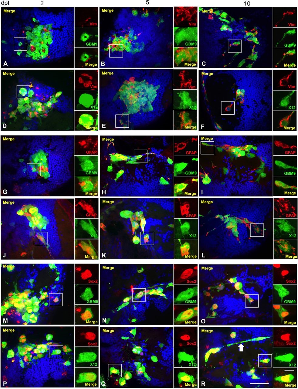
Fig. 6
GBM9 and X12 tumors contain a combination of differentiated cells and stem cells. Confocal images of GBM9 and X12 on 2 (A,D,G,J,M,P), 5 (B,E,H,K,N,Q) and 10 (C,F,I,L,O,R) dpt transverse cryosections. (A-C) GBM9 (green), DAPI (blue) and vimentin (red) at 100×. (D-F) X12 (green), DAPI (blue) and vimentin (red) at 100×. (G-I) GBM9 (green), DAPI (blue) and GFAP (red) at 100×. (J-L) X12 (green), DAPI (blue) and GFAP (red) at 100×. (M-O) GBM9 (green), DAPI (blue) and Sox2 (red) at 100×. (P-R) X12 (green), DAPI (blue) and Sox2 (red) at 100×. White boxes denote area magnified to the right of the image. White arrow in R points to a cell with a migratory morphology. n=5 animals per group; 90 total animals. Scale bar: 20µm for main panels and 5µm for insets.