
Image
|
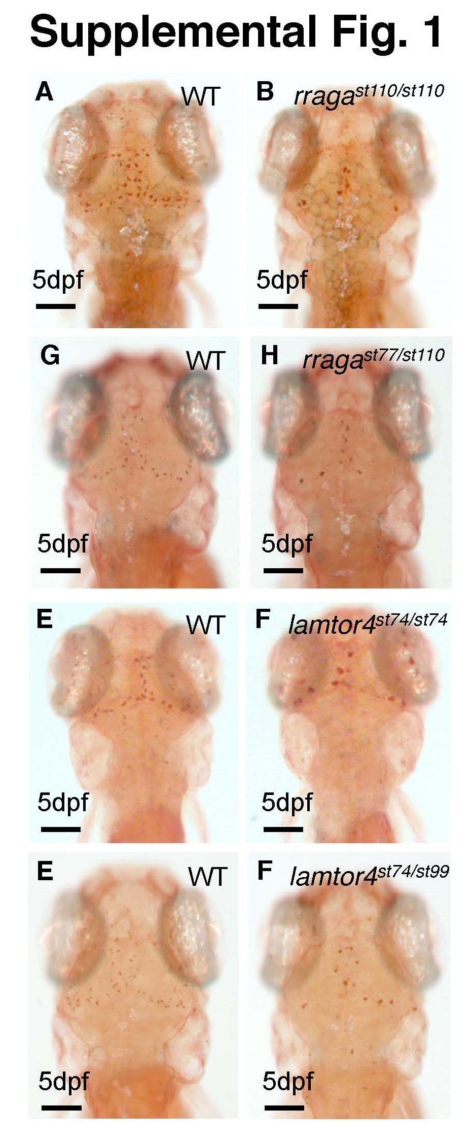
Figure Caption
Fig. S1
Related to Figure 1. Neutral red staining of rragast110/st110, rragast77/st110, lamtor4st74/st99, lamtor4st74/st99 mutants.
(A-H) Images of living zebrafish larvae of the indicated genotypes at 5 dpf after neutral red staining. Microglia were reduced to a similar extent in all mutants. Dorsal views, anterior to the top. All scale bars are 50µm. All larvae shown were genotyped by PCR after photography.
Acknowledgments
This image is the copyrighted work of the attributed author or publisher, and
ZFIN has permission only to display this image to its users.
Additional permissions should be obtained from the applicable author or publisher of the image.
Full text @ Cell Rep.