
|
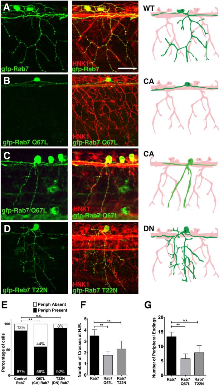
Fig. 2
Peripheral axon outgrowth and branching defects in CA-Rab7 expressing neurons. a-c Confocal projections of embryos with all RBs labeled with HNK-1 antibody (red) and individual RBs labeled with indicated Rab7 forms (green). Drawings at right highlight morphology of one neuron in green. a Individual RB neuron labeled with GFP-Rab7 showing central axons extending anteriorly and posteriorly from the cell body, and peripheral axon branching in the skin. b Lack of peripheral axon outgrowth in GFP-Rab7 Q67L (CA) expressing cell. c Reduced peripheral branching in GFP-Rab7 Q67L (CA) expressing cell. d Normal central outgrowth and peripheral branching in GFP-Rab7 T22N (DN) mutant expressing RB cell. e Expression of CA-Rab7, but not DN-Rab7, mutant construct increases percentage of RB neurons that do not extend a peripheral axon. n = 31 cells in Rab7 control, 30 cells in CA-Rab7, and 14 cells in DN-Rab7, **p = 0.006, Chi-Square test. f Number of peripheral axons that cross horizontal myoseptum (H.M.) is reduced in CA-Rab7 expressing cells. CA-Rab7: n = 9 cells; wildtype Rab7 control: n = 12 cells. **p = 0.009, paired t-test. g Number of peripheral axon endings in individually labeled neurons **p = 0.005, Unpaired two tailed t-test. Wildtype Rab7: 31 cells in 19 embryos; Rab7 Q67L: 30 cells in 15 embryos; Rab7 T22N: 14 cells in 9 embryos. All views anterior to the left. Scale bar = 40 µm