
|
Fig. 1
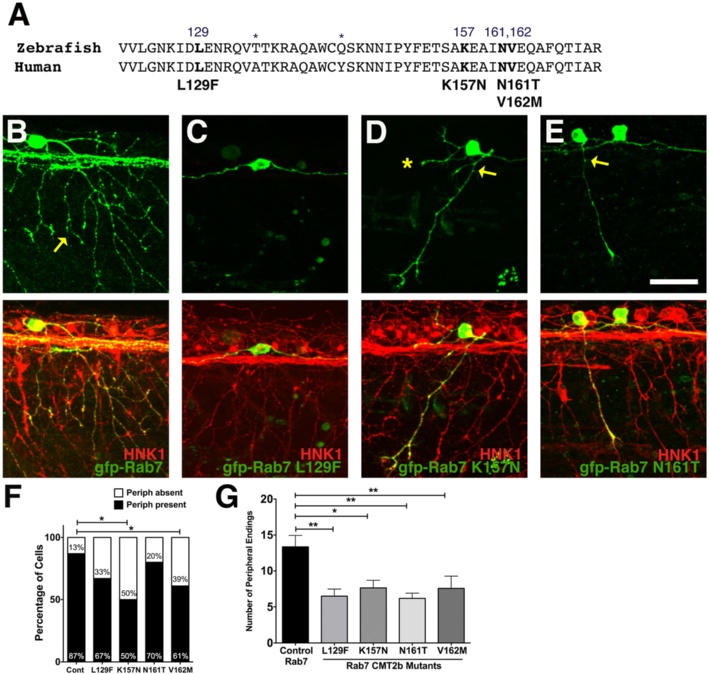
Decreased peripheral axon branching in sensory neurons expressing CMT2b Rab7 mutants. a Comparison of zebrafish and human Rab7 protein sequence in the region of CMT2b mutations. Differences in amino acid sequence indicated by asterisks. Amino acid residues associated with CMT2b are bolded. b-e Confocal images of individual RB neurons expressing wildtype GFP-Rab7 (b) or CMT2b GFP-Rab7 mutants (c) in embryos with all RB neurons labeled with HNK-1 antibody (red). Anterior to the left. b wildtype Rab7 expressing cell shows wide arborization of peripheral axons (yellow arrow). c Lack of peripheral axon outgrowth in Rab7 L129F expressing cell. d, e Reduced peripheral branching (yellow arrow) in Rab7 K157N (D) and Rab7 N161T (E) expressing cells. Asterisk = missing central axon. f Quantification of number of neurons extending peripheral axons. Cont = wildtype Rab7: n = 31 cells in 19 embryos; Rab7 L129F: n = 33 cells in 24 embryos, p = 0.08; Rab7 K157N: n = 10 cells in 10 embryos, *p = 0.03; Rab7 N161T: n = 15 cells in 14 embryos, p = 0.6; Rab7 V162M: n = 23 cells in 18 embryos, *p = 0.05. Fisher’s exact tests. g Quantification of peripheral branch endings in CMT2b-associated Rab7 mutations shows a significant decrease in branching. Wildtype Rab7: n = 20 cells in 15 embryos; Rab7 L129F: n = 21 cells in 18 embryos; Rab7 K157N: n = 11 cells in 7 embryos; Rab7 N161T: n = 12 cells in 10 embryos; Rab7 V162M: n = 16 cells in 14 embryos. *p = 0.03, **p < 0.01; Unpaired two-tailed t-test. Scale bar = 40 µm