
|
Fig. S3
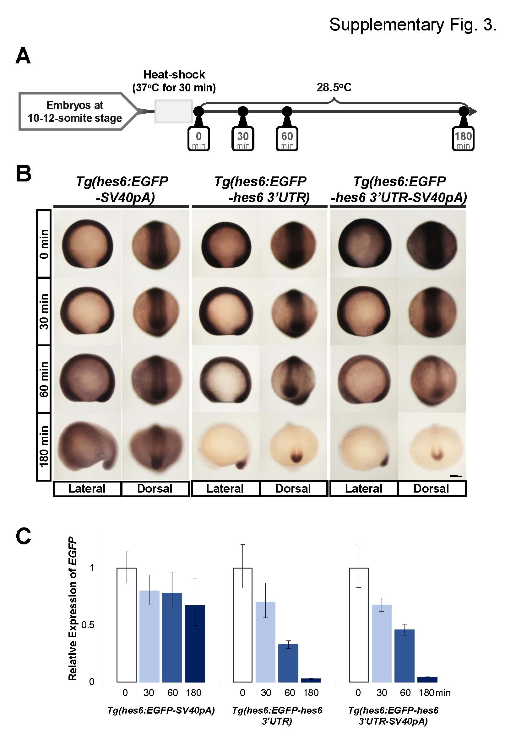
Comparisons of mRNA decay between EGFP-SV40pA, EGFP-hes6 3′UTR,, and EGFP hes6 3′UTR-SV40pA mRNAs induced by the heat-shock treatments on the transgenic embryos. (A) Schematic representation of heat-shock experiments. In the transgenic lines used in this study, the heat shock promoter, hsp70l, was used as a basal promoter for the reporter gene (Fig. 2A, 6A, and 7A), and the reporter gene could be ubiquitously induced by heat-shock treatments. To induce the EGFP mRNAs, heat-shock treatment was carried out for 30 min at 37°C on 10–12-somite stage embryos. After heat-shock treatment, embryos were incubated at 28.5°C and analyzed at 0 min, 30 min, 60 min, and 180 min. (B) Representative images of EGFP mRNA expression patterns induced by heat-shock treatment. Whole-mount in situ hybridization was performed using EGFP antisense probe. All images were taken at the same magnification. Scale bar, 200 µm. (C) Quantitative PCR to examine the decay of EGFP mRNA induced by heat-shock treatment. EGFP mRNA levels were normalized with zebrafish EF1α levels. Error bar represents the standard deviation.
Reprinted from Developmental Biology, 409(2), Kawamura, A., Ovara, H., Ooka, Y., Kinoshita, H., Hoshikawa, M., Nakajo, K., Yokota, D., Fujino, Y., Higashijima, S.I., Takada, S., Yamasu, K., Posterior-anterior gradient of zebrafish hes6 expression in the presomitic mesoderm is established by the combinatorial functions of the downstream enhancer and 3'UTR, 543-54, Copyright (2016) with permission from Elsevier. Full text @ Dev. Biol.