
|
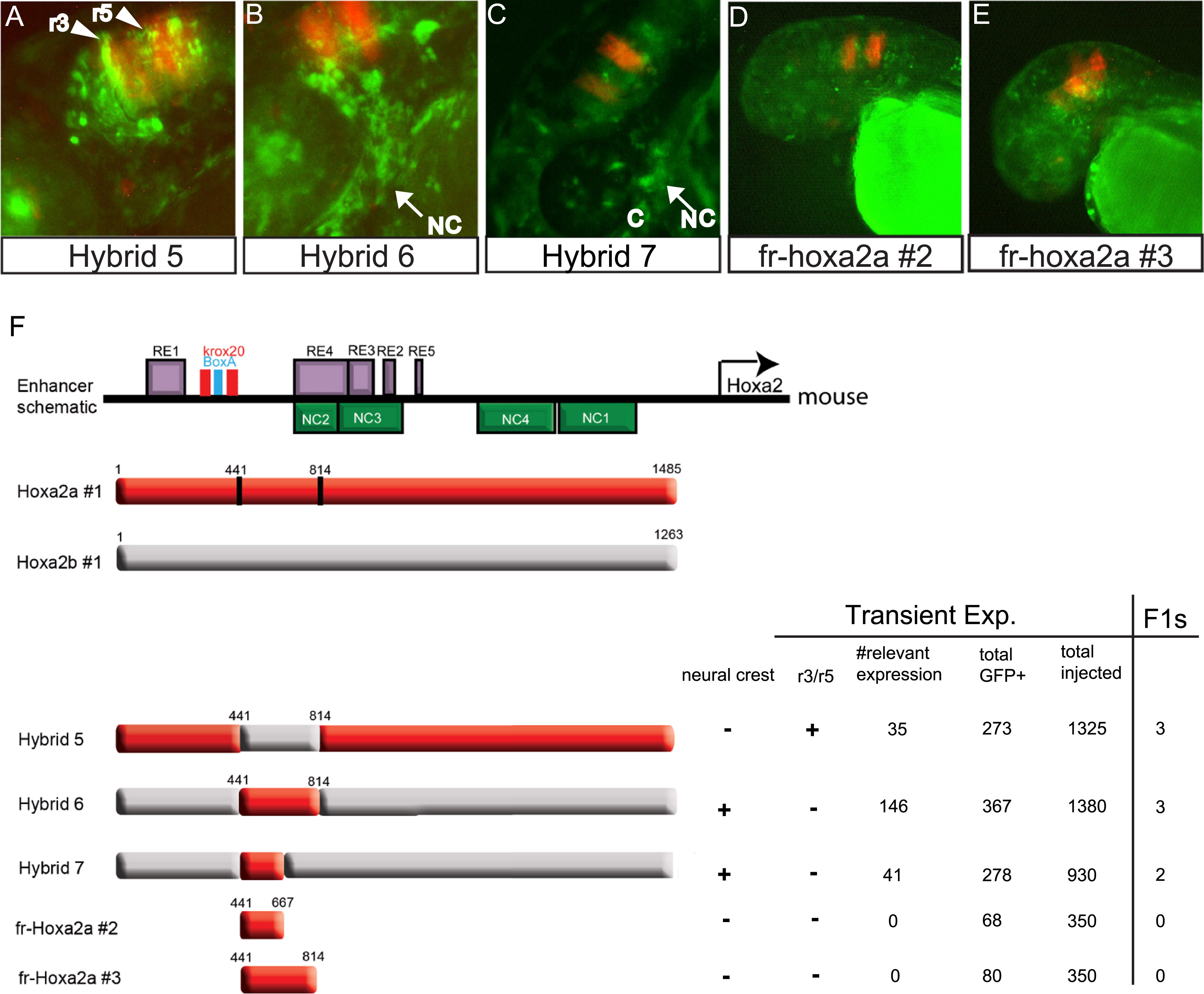
Fig. 5
Transgenic analysis of chimeric fugu hoxa2a/hoxa2b enhancers in zebrafish embryos define NC5 as a cis-element important in neural crest expression. (A-E) GFP reporter expression mediated by Hybrids 5-7 and isolated regions of hoxa2a (fr-Hoxa2a #2 and fr-Hoxa2a #3) in neural crest cells (NC) in pharyngeal arch tissue (arrows) or rhombomeres 3 and 5 (arrowheads) in 48hpf zebrafish embryos. In A-D: red=RFP expression in r3 and r5 driven by a control Krox20 enhancer; green=GFP expression. (F) Drawing of the hybrid constructs mapping the portion of the hoxa2a and hoxa2b enhancers in each hybrid or the small isolated regions compared to the annotated mouse Hoxa2 enhancer at top. The purple box= rhombomere elements; green box=neural crest elements; red=hoxa2a enhancer sequence; gray=hoxa2b enhancer sequence. The table at the right of each construct describes the F0 transgenic expression data marking: presence (+) or absence () of GFP expression in neural crest or r3/r5; # of embryos with expression in neural crest and/or r3/5 (#relevant expression); # of embryos with any GFP expression; total # embryos injected. The number of stable transgenic lines created (F1s) are noted, far right.
Reprinted from Developmental Biology, 409(2), McEllin, J.A., Alexander, T.B., Tümpel, S., Wiedemann, L.M., Krumlauf, R., Analyses of fugu hoxa2 genes provide evidence for subfunctionalization of neural crest cell and rhombomere cis-regulatory modules during vertebrate evolution, 530-42, Copyright (2016) with permission from Elsevier. Full text @ Dev. Biol.