
Fig. 2
- ID
- ZDB-IMAGE-160304-11
- Publication
- Diotel et al., 2016 - Mapping of Brain lipid binding protein (Blbp) in the brain of adult zebrafish, co-expression with aromatase B and links with proliferation
- All Figures
- Figures for Diotel et al., 2016
|
Fig. 2
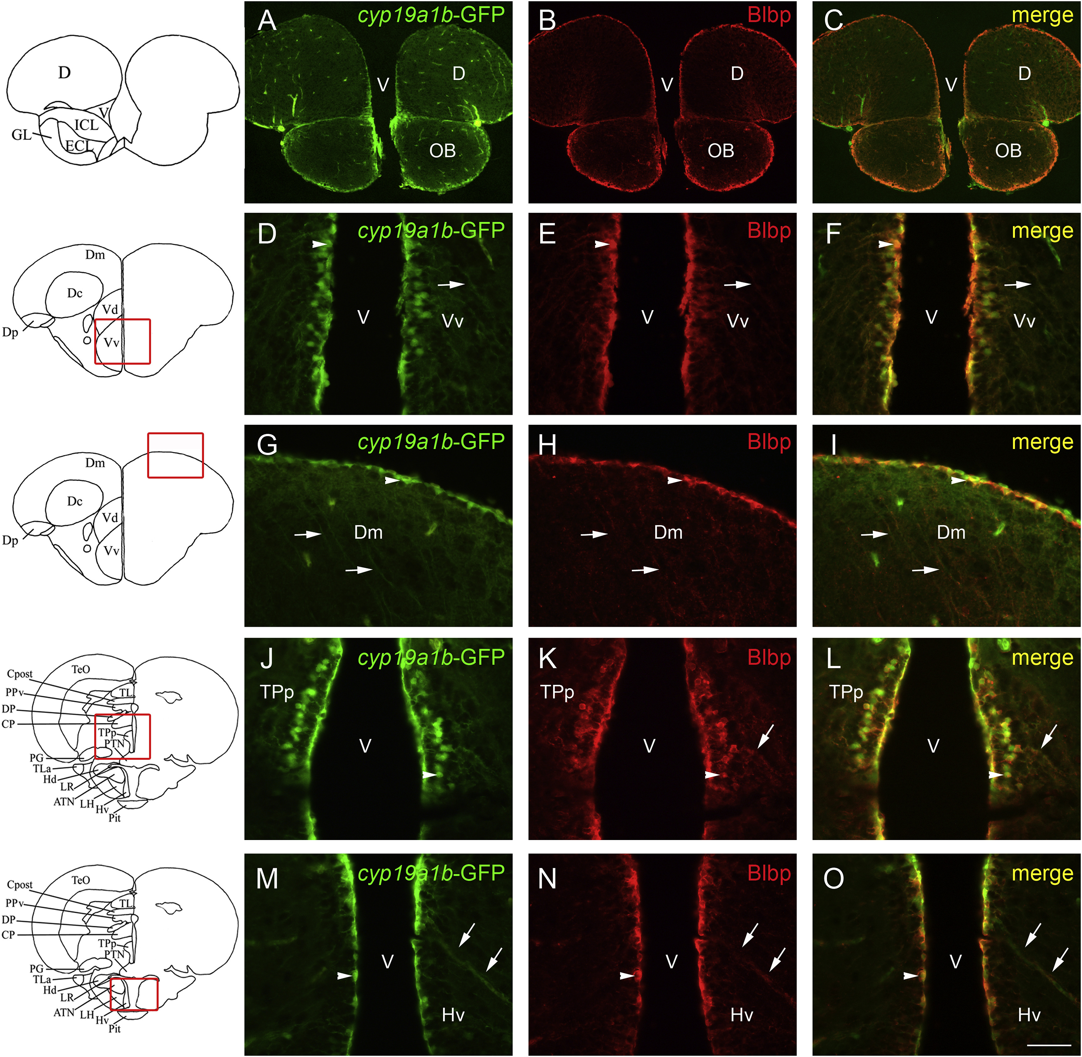
cyp19a1b-GFP and Blbp co-expression in the brain of adult zebrafish. A-O: Blbp (red) immunohistochemistry on cyp19a1b-GFP (green) transgenic zebrafish line shows that most radial glial cells co-express Blbp and GFP. Co-expression is obvious at the junction between the telencephalon and the olfactory bulbs (A-C), in the subpallium (D-F), and in the pallium (G-I). In the anterior part of the periventricular nucleus of the posterior tuberculum, GFP and Blbp are co-expressed (J-L) as well as in the anterior region of the hypothalamus (M-O). Arrows point to radial glial processes co-expressing GFP and Blbp, while arrowheads highlight co-expression in soma. Bar: 25 µm (D-O); 100 µm (A-C).
Reprinted from Gene expression patterns : GEP, 20(1), Diotel, N., Vaillant, C., Kah, O., Pellegrini, E., Mapping of Brain lipid binding protein (Blbp) in the brain of adult zebrafish, co-expression with aromatase B and links with proliferation, 42-54, Copyright (2016) with permission from Elsevier. Full text @ Gene Expr. Patterns