
|
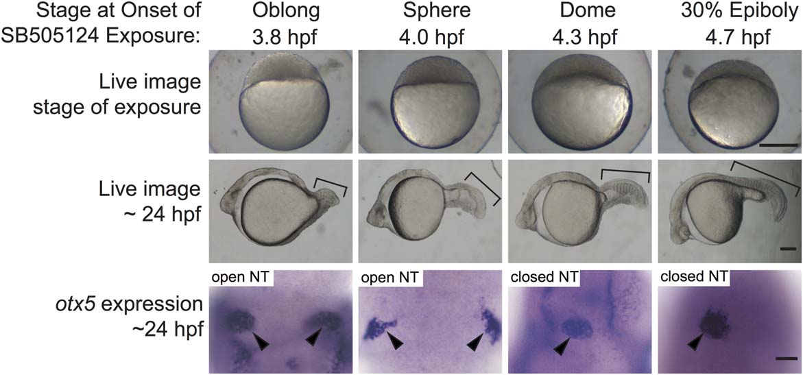
Fig. 1
Failure of neural tube closure in embryos treated with the Nodal inhibitor SB505124 for 20 min during mid blastula stages (3.8 and 4.0 hpf). Embryos treated with 75 µM SB505124 at all four stages of development have cyclopic eyes. The severity of the defects in mesendodermal/mesodermal derivatives, such as somites (brackets) decreases as the treatments move later in development. Embryos treated for 20 min at 3.8 and 4.0 hpf have open neural tubes (open NT), as evidenced by the two domains of pineal precursors (arrowheads). In contrast, embryos treated at 4.3 and 4.7 hpf have an oval shaped pineal anlage (arrowheads) indicating a closed anterior neural tube (closed NT). Top row: lateral views, animal pole to the top, scale bar: 250 µm. Second row: lateral views anterior to the left, scale bar: 50 µm. Third row: dorsal views anterior to the top. Scale bar: 50 µm. Refer to Table 1 for quantitative data.